Showing posts with label MAP. Show all posts

Sunday, December 7, 2014

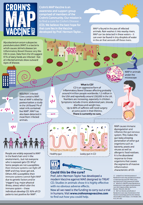
Infographic Snapshot of the Lifecycle of #MAP in People w/ #Crohns

Great infographic image of the life cycle of MAP.  


The part of this picture that should jump out at you is the statistic on the bottom left. citation # 3

92% !!!....... this is madness!  This isn't new news people.  So why doesn't anyone know about MAP and it's relation to Crohn's?  I'm not talking about lay persons, but people in the profession of helping people get better who are living with this disease.  They don't acknowledge it.  The medical community flat out ignores the science and the 30 years or more of research that exists.   People need to take charge of their life and health and start fighting to know something.  Ask questions and demand an answer is where I'd start.  


Click link below to view image larger from original source:

Saturday, September 20, 2014

Crohn's - MAP Bacteria Connection *Video*

Informative 3 min. YouTube video to raise awareness about the Crohn's-MAP bacteria connection.  How humans are exposed and vulnerable to MAP - There are now ways to test for MAP.   
I also posted the links that were provided on the YouTube page under the video for further reading/info.


- The Crohn’s MAP Vaccine
A modern therapeutic Vaccine against MAP has been created. Preliminary studies in animals have shown it to be safe and effective. Next, a trial in humans is needed to take the vaccine from lab to clinic.
Professor John Hermon-Taylor, King's College London
http://crohnsmapvaccine.com

Crohn's Disease Info Center:
http://www.shafran.net/crohn/TrialRes...

Providing the highest quality medical care, Shafran Gastroenterology Center, IBD Center-of-Excellence in Florida:
http://www.shafran.net/center/

CDD The Centre for Digestive Diseases, Crohn's Disease:
http://www.cdd.com.au/pages/disease_i...

Efficacy and Safety of Anti-MAP Therapy in Adult Crohn's Disease (MAPUS), RHB-104:
http://www.clinicaltrials.gov/show/NC...

RHB-104 is a proprietary antibiotics drug combination for treatment of Crohn’s disease:
http://www.redhillbio.com/product-pip...

Thursday, February 6, 2014

Is It Possible to Cure Crohn's Disease? I Think So

A writer for Psychology Today, contributes the following article (and 2 more that I will post after this article) pertaining to something that I have read up on and firmly believe is the culprit to Crohn's disease.  Everyone should read this information and remember it.  I think a lot of people have been made hopeless or skeptical in regards to Crohn's disease because people are told so many things that contradict that there will ever be a cure.  Doctor's have been telling people for decades now that there will never be a cure.
It's a new day people, get out of the rut of thinking in the past and open your mind to new developements and discoveries.  It's worth it and I am super duper hopeful.


Through luck, hard work, good fortune, perseverance, and wonderful doctors, I seem to be one of the few people in the world who can claim to be “cured” of Crohn’s Disease. I’ve told this story before, in several places, but I feel a need to get the information out to a more general audience, even though this is not a psychology story but a medical and scientific one. In September, 2005, I wrote a small article for the PCC SoundConsumer, a very small newspaper published by a local Seattle natural foods grocery store, about my experience with Crohn’s Disease. http://www.pccnaturalmarkets.com/sc/0509/sc0509-hw-crohns.htm For some reason, that little article went viral, and to this day I receive letters from people from all over the world asking for information about Crohn’s Disease and its probable connection with a specific microbe, mycobacterium avium subspecies paratuberculosis (MAP). I told the story again in a scientific journal, the Paratuberculosis Newsletter,http://www.paratuberculosis.info/web/images/newsletters/2012q2.pdfBut that journal is not highly visible. Since 8 years has gone by, and I have much more information now, I am going to put this on Psychology Today, for the benefit of all of those who suffer from Crohn’s or who love people who suffer from Crohn’s. I will have to tell this story in several segments, since Psych Today restricts individual posts to about 800 words. Please bear with me. I will be providing a lot of information, references, and information that is crucial to individual and public health.Single case reports are no longer the fashion in modern medicine. We all know that an n of 1 plus 1 plus 1 plus 1 ad infinitum equals nothing in terms of proving causality in “evidence based medicine”. Proving anything in the case of Crohn’s Disease is difficult. See this excellent review of this situation, Causality and gastrointestinal infections: Koch, Hill, and Crohn’s by Anne-Marie Lowe, Cedric P Yansouni, and Marcel A Behr, Lancet Infect Dis 2008:8: 720–26. You must understand that for scientists today, a single case report is virtually meaningless. However, my victory over Crohn’s Disease doesn’t prove anything by itself, but I think that it matters insofar as it suggests the desirability of pursuing a more detailed scientific inquiry. Furthermore, there are hundreds of thousands of people with Crohn’s, many of them children, who are being treated under a different paradigm, and with variable success. Moreover, I think many of those people got sick because they consumed contaminated food products without knowing it, sometimes years before the disease manifested itself. I think there is better way to treat Crohn’s, and better yet, to prevent it. In brief, I think I was “cured” of Crohn’s Disease with antibiotics directed at the eradication of Mycobacterium avium subspecies paratuberculosis.Background: MAP is a small bacteria in the same family as leprosy and tuberculosis. In 1894, two veterinarians, Dr. H.A. Johne and Dr. L. Frothingham, identified this organism in a cow with weight loss and poor milk production in Germany. It is an acid-fast bacterium, like TB and leprosy, but has other properties as well. The best place to read about MAP is on the web site www.johnes.org

Farmers rapidly learned that their cows could develop either bovine TB or what came to be known as Johne’s Disease. Both were serious and expensive for the farmers. However, for various reasons, everyone agreed that tuberculosis could be transmitted from cows to people, and many public health measures were put into place to stop bovine TB. However, to this day, the USDA and the IOE, the World Organization for Animal Health http://www.oie.int/ deny that MAP is a “zoonotic disease”, a disease that can be transmitted from animals to people. The ostensible reasons for this are that MAP cannot be visualized in human tissues with ordinary microscopes (while it is easy to see MAP in animal specimens); PCR studies and other DNA fingerprinting technologies yield ambiguous results; and nobody dares to try the experiment to prove Koch’s postulates, which would be to grow human MAP in culture, and then feed those germs to human infants. That would be immoral and ridiculous. For various reasons, scientists dispute the claim that MAP causes Crohn’s Disease because Koch’s postulates have not been fulfilled, even though nobody has ever grown M. Leprae (the germ that causes leprosy) in culture. It has to be grown in the foot pads of armadillos…. Koch’s postulates are not applied to viruses, prions, or other pathogens. Even though MAP comes very close to meeting Koch’s postulates, its pathogenicity is disputed. (There was even an experiment in the 1980s in which MAP was grown from human biopsy specimens, and then fed to baby goats. The goats got Johne’s Disease. But it was a small n…)
As a result, animals that test positive for MAP are not culled and eliminated from human food products. They can be sold for meat, into the food supply. Moreover, viable MAP can be cultured from milk, including pastuerized milk, around the world. MAP is a very stubborn bacteria, resistant to heat and chlorine. It thrives on biofilms, and has a persistent spore that can last for years. The incidence of Crohn’s Disease in the world has grown in proportion to the use of dairy products. For example, Japan had little Crohn’s 100 years ago, and now it is becoming a significant problem. When you go to the grocery store and consider buying raw milk products, remember that MAP is endemic to cows, sheep, and goats, and roughly 1% of pasteurized milk in the US contains viable MAP.

Thursday, December 19, 2013

Borody-His Innovative Discoveries on Digestive Disease - #FMT & #RHB104

This is another great article.  I knew that Dr. Borody, a doctor and professor out of Australia, developed FMT's (Fecal Matter Transplants) and was 1st to use the transplants to treat c-diff.  Interesting enough he is also considering FMT's for other digestive problems and even neurological conditions.  It will be interesting to see what illnesses are helped with this procedure.  

Here in the US doesn't even have a billing code for FMT's AND the FDA have only permitted transplants for difficult cases of c-diff, patients who do not respond to the antibiotic, that only around 30% of the patients respond to.  FMT's have a 90% recovery rate.  The question again is why not go with the treatment that has a higher success rate rather than lower.  Very sad that the US is so slow to adopt new and smarter treatments that will get people well.   I won't even go there today. It is an issue that probably raises my blood pressure if I think about it too long.  Not even joking.  

Anyway, after reading this article, I found out that Dr Borody also was the innovator for RHB-104 that is currently in the trial stages at this time. If you have Crohn's and don't know what RHB-104, read up on the treatment.  I have some blog posts that will give a good amount of information about the therapy.  It will make you smile and give you a boost of hope for something that may actually be able to eradicate Crohn's all together.  That is exciting!  

Professor Thomas Borody gets it!  He seems to understand the nature of digestive diseases very well.  He is certainly the man who is making some progress with developing real solutions to treat the actual condition and not just remedy the symptoms of a disease.  His name is one to remember and follow as he implements treatments that seem to be successful so far.  
Part of this article gives a little in depth look at his life and how he chose to dedicate his direction to gastrointestinal diseases.   

Note: I have underlined parts of the article that I felt were things to take note of.

He has the stomach for it | JPost | Israel News:

''Prof. Thomas Borody
Prof. Thomas Borody Photo: Courtesy
It’s amazing when in medicine, ideas that had seemed ridiculous – such as a chronic infection instead of an annoying boss causing ulcers – eventually become accepted truisms. Even though stools are considered dirty and something to get rid of, gastroenterologists have discovered that they can transplant feces from healthy donors into patients and very often relieve their chronic bacterial infections, irritable bowel disease (IBD), colitis, constipation and even some neurological conditions.

Prof. Thomas Borody, the founder and current medical director of the Center for Digestive Diseases (CDD) in Sydney, Australia, is a major innovator in these surprising treatments.

The CDD is considered a unique medical institution offering novel approaches in researching, diagnosing and treating gastrointestinal conditions. CDD offers a range of services in the day-procedure unit and houses a research and innovation department for conducting clinical trials in collaboration with universities, pharmaceutical companies and medical societies – all focusing on gastroenterology.


HE RECENTLY visited Israel and gave an interview to The Jerusalem Post about new treatments in the field. “This is my first visit to Israel,” said the world-renowned gastroenterologist.

“I’ve given a gastroenterology lecture to professionals at the Dan Hotel in Tel Aviv. I’ve also done some touring in Jerusalem, the Dead Sea and elsewhere – and I’ve already decided to come back.”

Borody was born in Krakow in 1950. My father was a Seventh-Day Adventist minister of religion,” said Borody. When he was 10, his parents decided to move to Australia, and his mother even wrote a book about their immigration experiences.

Inculcated to learn something that would “help mankind,” he and his siblings studied medicine. “I had thought of becoming a nuclear physicist, but it wasn’t to be,” he recalled. His sister is a general practitioner, his brother is an obstetrician/gynecologist and Thomas decided after getting his MD to specialize in the stomach, intestines and other internal organs.

He was inspired to pick his specialty largely because he worked under Dr. David burns, who was “a nice guy and did gastroscopes, which just came into being, in color.

Gastroenterologists spend much of their time performing colonoscopies (using endoscopes with tiny cameras in them to examine the large intestine for polyps or tumors and other irregularities) and gastroscopies (an endoscope is inserted into the mouth and through the esophagus to the stomach to get a view of the upper part of the gastro system). Since they have to fast and clean out their tubes, the gastro practice usually begins early in the morning.

AS FOR ulcers, it was two Australians who discovered in 1979 that they were not the result of stomach acid, spicy food and stress but a bacterium – Helicobacter pylori (H.

pylori) – that caused a chronic stomach infection. Dr. Barry James Marshall and pathologist Dr. John Robin Warren received the Nobel Prize in Medicine in 2005 for their breakthrough in discovering what causes ulcers.

It had been thought by the medical establishment for many years that no bacteria could survive in stomach acid, so that the painful condition must be caused by something else. But the two Australians found it is a very “clever” pathogen that can hide in the mucous lining protected from the acid, so they could remain alive even for decades.

Borody reasoned that if it was due to an infection, something could be done to cure it. So he and his colleagues developed in 1984 a triple “cocktail” of drugs – bismuth, tetracycline and flagyl – to treat and eliminate stomach ulcers. “We went through about 36 different combinations of drugs, and the best were these three.” When ulcer patients come to thank him that their ulcers have disappeared, he said, he feels “great satisfaction.” But despite proof that this healed the stomach lining, it took many years until the doctors were persuaded and accepted the triple therapy. The bacteria return only very rarely, and if it’s caught early, a recurrence can also be treated.

Today, one of the companies that manufactures the cocktail is RedHill Biopharma, an Israeli company. During his visit here, he met with officials of the company, which is based in Tel Aviv. Borody is an unpaid member of the RedHill’s advisory board.

There are other gastroenterological conditions, he continued, that is through to result from lifestyle that in fact result from chronic inflammation – among them ulcerative colitis and Crohn’s disease, which cause much suffering to many people around the world.

“After practicing for a while, I fell into research and spent year in the Solomon Islands [in Oceania lying to the east of Papua New Guinea] doing tropical med. I was more involved in leprosy and tuberculosis, which also involve bacteria that need a cocktail of drugs to treat it. This led to my getting interested in Crohn’s disease and irritable bowel disease.”

Borody spent three years researching at the Mayo Clinic.

NAMED AFTER Dr. Burrill Crohn, who first described the disease 80 years ago, Crohn’s disease is a chronic inflammatory condition of the gastrointestinal tract. It belongs to a group of conditions known as inflammatory bowel diseases (but is not the same as ulcerative colitis, which is another type of IBD. The symptoms of these two conditions are quite similar, but they affect different areas of the gastrointestinal tract. Crohn’s most often affects the end of the small bowel and the beginning of the colon, but it may affect any part of the gastrointestinal tract, from the mouth to the anus. Ulcerative colitis is restricted to the colon, also called the large intestine.

Among the symptoms of Crohn’s, which can be very debilitating and limiting, are persistent diarrhea; rectal bleeding; the urgent need to move bowels; abdominal cramps and pain; constipation; and the sensation of incomplete. It can be accompanied by loss of appetite; weigh loss; fatigue; night sweats; and the loss of a normal menstrual cycle in women. It is not a genetic condition, although some used to think so because of cross-infection in the family. It was also thought to be an autoimmune disease in which the autoimmune system, which is supposed to protect the body against infections, becomes aggressive and uncontrollable and attacks the body itself.
“Crohn’s is a more difficult disease to treat,” noted Borody. Because the bacterial infection develops very slowly. The bacteria involved are the most slowly dividing bacteria known to man. “This process can take 15 months, so medications have to be taken for a long time. It’s a difficult bug to destroy until the inflammatory condition calms down.”

Twenty years ago, Borody was reading The Lancet medical journal. “There as a published reader’s letter I came across that mentioned that a person was treated for tuberculosis, and his Crohn’s also improved. In Crohn’s, you see granulomas in the chest that are surrounded by epithelial cells. It looks like TB, but it’s Crohn’s. So I thought it could be treated with a new type of anti-tuberculosis agents. We started 12 Crohn’s on these drugs and it takes many weeks to get better. The longest time a patient has been on these drugs is 19 years.”

Now, a new, experimental drug for Crohn’s developed and patented by Red- Hill Biopharma now undergoing Phase III studies is called RHB-104. “Just as tuberculosis and HIV requires taken a drug cocktail to fight them, Crohn’s does too. If you give too-few medications, resistance to them can develop.

RHB-104 is a proprietary and potentially grandbreaking combination antibiotic therapy in oral pill form, with potent intracellular, antimycobacterial and anti-inflammatory properties, RedHill explains.


“The drug is based on increasing evidence supporting the hypothesis that Crohn’s disease is caused by an infection of the Mycobacterium avium subspecies named paratuberculosis in susceptible patients rather than being an autoimmune disease.

Borody notes that the triple drug combination that he invented for Crohn’s have been on the market for years, but the combination he suggested is new. “There are now clinical trials in 40 countries around the world. There are an estimated two or three million Crohn’s patients globally.

The phenomenon is grow- ing in places like China, Hongkong and Japan.”

When the US Food and Drug Administration approves the drug cocktail, “it will explode on the market. There is much need for such a treatment. It is legal to combine separate prescription drugs, but this will be a single capsule.”

The food supply around the world is tainted because of cattle and sheep getting it, both their meat and their milk. So if such animals are infected, some of it can get to people if not properly prepared, said Borody
. The milk has to be pasteurized at very high temperatures but that changes the quality.

It is difficult to detect the bacteria in meat. About 60% of America herds catch it, and they have gotten to poultry as well.

AS FOR stool transplants – known scientifically as fecal microbiota transplantation (FMT), Borody is very enthusiastic. “I read a scientific paper from way back in 1958 about a patient being given feces for mild colitis caused by the Clostridium difficile bacteria. But it didn’t go further.” The bacteria produce anything from diarrhea to Pseudomembranous colitis. More virulent strains have been developing in the last 15 years because the overuse of antibiotics caused resistance to them. In the U.S alone, there are some three million new cases of such infections each year; some of them can even be fatal.

In FMT, a donor with healthy and beneficial bacteria (probiotica) in his gastrointestinal system gives some 150 cc. of feces that are purified and introduced by enema into the recipient. A healthy growth of bacteria in the gastroenterological system is restored. A randomized study on FMT published in the New England Journal of Medicine last January showed a 94% cure rate of pseudomembranous colitis caused by Clostridium difficile, compared to just 31% with the antibiotic vancomycin. The study was stopped prematurely as it was considered unethical not to offer the FMT to all participants of the study due to the outstanding results.

Wednesday, December 18, 2013

Identifying & Destroying the Cause of Crohn's Disease #ibd #MAP

Encouraging and hopeful article!  


'UCF College of Medicine professor Dr. Saleh Naser soon will participate in a clinical trial to test whether a new antibiotic therapy acquired by RedHill Biopharma can be used to treat Crohn’s disease patients.
The FDA-approved phase III trial is expected to commence within weeks by RedHill Biopharma, which licensed Naser’s DNA technology for detecting Mycobacterium avium subspecies paratuberculosis, known as MAP. It is believed to be associated with Crohn’s disease. RedHill Biopharma developed the anti-MAP antibiotic regimen known as RHB 104. Crohn’s disease is a chronic inflammatory disease of the gastrointestinal tract characterized by cramping and diarrhea.
Naser developed and patented a way to detect MAP from milk, blood and tissue clinical samples. The bacterium is known to cause inflammation in the intestines of cows. It is also linked to Crohn’s disease, although its role has been debated for more than a century. Naser believes MAP is an underlying cause of the disease.
“Crohn’s disease affects more than 750,000 Americans, yet traditional treatments only address the symptoms of inflammation and not the cause,” Naser said. “I have seen case studies where patients’ lives have been restored following treatment, which removes MAP. I have high hopes that this clinical trial may lead to finding a cure.”
RedHill will be enrolling 240 subjects from the United States, Canada and Israel in this double blind clinical trial in which blood and intestinal biopsy specimens from Crohn’s patients will be tested for MAP before, during and following the one-year treatment with the antibiotic RHB 104.
“Since we acquired the license to Dr. Saleh Naser’s MAP detection technique in 2011, we have had an excellent collaboration with UCF,” said RedHill’s CEO Dror Ben-Asher. “The UCF team of researchers… is at the forefront of global academic research on MAP and its detection.”
Naser is looking forward to the trial and hopes this will end the academic debate regarding MAP and Crohn’s disease.
“I am ecstatic to be part of a team, which will help determine whether or not MAP is associated with Crohn’s disease; certainly a final answer to a one hundred-year old controversy,” Naser said.
Naser joined UCF in 1995 and has been a faculty member in the medical college since its foundation. He teaches clinical chemistry and infectious processes in the Burnett School of Biomedical Sciences within the medical school. He also serves as the graduate coordinator for three masters programs in the College of Medicine.

Sunday, October 6, 2013

REDHILL BIOPHARM - News -> RHB-104 - Possible Breakthrough Treatment for Crohn's Disease


I get so excited when I read about this possible treatment, RHB-104, for Crohn's!  I've been watching this drug for over a year now.  This is a  treatment that if approved by the FDA, I will be jumping on it as soon as it becomes available.  No doubt!  This is the kind of treatment we need.  Everything else just treats symptoms.
So, why is this so exciting?  If you read current research about what causes Crohn's disease, you will see that researchers are leaning away from considering Crohn's disease an autoimmune condition.  They have found that there is an underlying cause for the immune response to activate.  Crohn's is said to be caused by a combo of 3 factors - Immune system abnormalities, environmental influences and bacteria.  I just wiki'd Crohn's disease and this is what is said about what is believed to be the cause - "Crohn's disease is caused by interactions between environmental, immunological and bacterial factors in genetically susceptible individuals.[4][5][6] This results in a chronic inflammatory disorder, in which the body's immune system attacks the gastrointestinal tract possibly directed at microbial antigens.[5][7] While Crohn's is an immune related disease, it does not appear to be an autoimmune disease (in that the immune system is not being triggered by the body itself)."
What this means is that people that have Crohn's disease do not have the immune system capabilities to destroy a form of bacteria that have been introduced into the body at some point in time.  As research has indicated, a large number of people with Crohn's disease happen to have the MAP bacteria in their body somewhere.  The exact location of the bacteria is not known at this time.  Who cares where it is, as long as a treatment is available that will destroy it is my opinion.
See why this makes me super hopeful?  If Crohn's is caused by this MAP bacteria & if this new combination antibiotic eradicates the MAP bacteria from people with Crohn's disease, this could be the answer to the Crohn's disease epidemic!  Can someone say CURE :)
Wouldn't that be glorious?  

Clinical trial for a new antibiotic therapy for Crohn’s treatment started:
Crohn’s disease is an inflammatory bowel disease which may affect gastrointestinal tract from mouth to anus, and has a wide variety of symptoms. Mycobacterium avium subspecies paratuberculosis (MAP) is believed to be associated with Cronh’s disease. Professor Dr. Saleh Naser, UFC College of Medicine, believes that MAP is the cause of the disease. He said, "Crohn's disease affects more than 750,000 Americans, yet traditional treatments only address the symptoms of inflammation and not the cause." He further added, "I have seen case studies where patients' lives have been restored following treatment, which removes MAP. I have high hopes that this clinical trial may lead to finding a cure." Dr. Saleh Naser will soon conduct clinical trials on a new antibiotic therapy acquired by RedHill Biopharma for curing the disease. 240 participants will be recruited for this double blind clinical trial.
http://health-beauty-2468.blogspot.com/2013/09/clinical-trial-for-new-antibiotic.html


RedHill Biopharma to initiate phase III trial of RHB 104 in Crohn's disease patients
UCF College of Medicine professor Dr. Saleh Naser soon will participate in a clinical trial to test whether a new antibiotic therapy acquired by RedHill Biopharma can be used to treat Crohn's disease patients.
The FDA-approved phase III trial is expected to commence within weeks by RedHill Biopharma, which licensed Naser's DNA technology for detecting Mycobacterium aviumsubspecies paratuberculosis, known as MAP. It is believed to be associated with Crohn's disease. RedHill Biopharma developed the anti-MAP antibiotic regimen known as RHB 104. Crohn's disease is a chronic inflammatory disease of the gastrointestinal tract characterized by cramping and diarrhea.
Naser developed and patented a way to detect MAP from milk, blood and tissue clinical samples. The bacterium is known to cause inflammation in the intestines of cows. It is also linked to Crohn's disease, although its role has been debated for more than a century. Naser believes MAP is an underlying cause of the disease.
"Crohn's disease affects more than 750,000 Americans, yet traditional treatments only address the symptoms of inflammation and not the cause," Naser said. "I have seen case studies where patients' lives have been restored following treatment, which removes MAP. I have high hopes that this clinical trial may lead to finding a cure."
RedHill will be enrolling 240 subjects from the United States, Canada and Israel in this double blind clinical trial in which blood and intestinal biopsy specimens from Crohn's patients will be tested for MAP before, during and following the one-year treatment with the antibiotic RHB 104.
"Since we acquired the license to Dr. Saleh Naser's MAP detection technique in 2011, we have had an excellent collaboration with UCF," said RedHill's CEO Dror Ben-Asher. "The UCF team of researchers- is at the forefront of global academic research on MAP and its detection."
Naser is looking forward to the trial and hopes this will end the academic debate regarding MAP and Crohn's disease.
"I am ecstatic to be part of a team, which will help determine whether or not MAP is associated with Crohn's disease; certainly a final answer to a one hundred-year old controversy," Naser said.
http://www.news-medical.net/news/20130918/RedHill-Biopharma-to-initiate-phase-III-trial-of-RHB-104-in-Crohns-disease-patients.aspx


RedHill Readying Phase III Trials In Crohn's

With a PDUFA target date of Feb. 3, 2014 for its migraine drug, RHB-103, and NDA filing planned for the first quarter of 2014 for the anti-emetic drug, RHB-102, RedHill Biopharma (RDHL) is now focusing its efforts on its flagship programs: RHB-104 for Crohn's disease and RHB-105 for Helicobacter pylori (H. pylori).
RedHill is preparing to begin a potential groundbreaking Phase III study in the current quarter in North America and Israel (the MAP U.S. Study) of Crohn's disease, using a novel patent-protected formulation that combines three antibiotic ingredients in a single capsule, and is planning and preparing a parallel Phase III study in Europe (the MAP Europe Study).
"Following the discovery of the link between H. pylori bacterium and peptic ulcers, there is a growing body of evidence supporting the proposition that Crohn's disease and other so-called autoimmune diseases, such as multiple sclerosis, are linked to infections," chief business officer Guy Goldberg says in an interview with BioTuesdays.com, referring to emerging scientific evidence that the microbiome, or the trillions of intestinal microbes, plays a major role in health and disease.
"There's a paradigm shift of viewing the body not as a single organism but as a collection of organisms, with the microbiome as the habitat within the body," he adds.
In 2005, researchers in Australia won the Nobel Prize for identifying H. pylori bacterium and the role it plays in causing peptic ulcers. Stress and lifestyle were long believed to be the primary cause of intestinal ulcers. Fellow Australian, Prof. Thomas Borody, who is now a member of RedHill's advisory board, developed the first antibiotic treatment for ulcers.
RedHill's two lead drug candidates - RHB-104, for Crohn's disease and multiple sclerosis (MS), and RHB-105, forH. pylori - were built on the success of Prof. Borody's approaches to gastrointestinal tract diseases and infections.
"RHB-104 is the biggest program in our pipeline," Mr. Goldberg says. "To the best of our knowledge, we are the only company with a Phase III combination antibiotic approach for treating Crohn's disease, even though there is a lot of academic research connecting Crohn's to a bacterial infection."
RedHill is currently advancing six clinical programs; two of which are expected to be reviewed by the FDA during 2014, and three that are entering pivotal clinical studies.
In a research report last month, Bioassociate Innovative Consulting reiterated its "buy" rating and price target of $14.91 per RedHill American Depositary Share, "given the multiple milestones approaching and the company being on-track with the clinical programs' timelines."
Crohn's is a severe inflammatory disease in the GI tract. Existing drugs, such as Remicade, treat the inflammatory symptoms of the disease by suppressing the immune system. However, they are widely considered to have a poor safety profile and limited long-term efficacy. "There clearly is a strong unmet medical need for a better alternative for Crohn's patients," Mr. Goldberg contends. The global market to treat Crohn's exceeded $3.5-billion in 2012.
Phase II Study Pictures in Crohn
(Click to enlarge)
Phase II Study Pictures in Crohn's Patients (Borody et al (2002), Digest Liver Dis 34:29-38)
He explains that RHB-104 is a novel and proprietary combination of three approved antibiotic ingredients - clarithromycin, clofazimine and rifabutin - targeting Mycobacterium Avium Paratuberculosis (MAP). Crohn's patients are believed to have seven times greater likelihood of being MAP-positive than non-Crohn's patients.
The MAP U.S. Study will enroll 240 moderately-to-severely active Crohn's patients at 50 sites in North America and Israel. The primary endpoint is the state of remission at week 26. The study will also examine safety, the maintenance of remission through week 52, efficacy outcome measures in relation to the presence of MAP infection and other secondary endpoints.
If the study is successful, RedHill may submit a new drug application to the FDA in 2015. RHB-104 previously received orphan drug status from the FDA for pediatric use. Mr. Goldberg says discussions are underway with European regulators to begin a second Phase III study with RBH-104 in Europe - the MAP Europe Study.
RedHill also is developing RHB-104 to treat patients with relapsing remitting MS. In June, the company commenced a Phase IIa proof-of-concept trial in Israel. "This is the first time that this type of antibiotic therapy will be tested on MS patients in a Phase II clinical trial," Mr. Goldberg contends.
RedHill's second lead program is RHB-105 in development to treat H. pylori bacteria, which plays an important role in gastritis, peptic ulcers and gastric cancer. Mr. Goldberg points out that H. pylori is increasingly developing resistance to treatment with clarithromycin and metronidazole, and that standard therapy fails in up to 30% to 40% of patients who continue to remain H. pylori-positive.
Phase IIa study in 2005 by Prof. Borody in Australia demonstrated over 90% eradication of the bacteria in 130 patients who had previously failed standard therapy with clarithromycin. Approximately three millionH. pylori-infected patients are treated annually in the U.S. market, resulting in a potential market estimated at $1-billion to $1.5-billion.
Mr. Goldberg explains that RHB-105 is a novel all-in-one combination of two antibiotics - rifabutin and amoxicillin - and a proton pump inhibitor, omeprazole. RedHill plans to begin a Phase II/3 clinical trial with RHB-105 during the quarter, he says, adding that a successful study could lead to an NDA filing in 2014.
Wednesday, May 1, 2013

Researchers Find Genetic Link Specific in Identifying Johne's Disease (Disease caused by MAP) in Cattle

John's the man!!!
Ewwww  think about it for a second.  $220 million each year is spent on sick cows that are affected by MAP bacteria (bacteria that causes Johnes Disease).  How many cows are we talking about here?? A LOT! No wonder the # of people with Crohn's Disease is quickly increasing.  All the freaking cows milk is tainted.  

Will vaccinating cattle protect them from such a nasty tough as steel bacteria like MAP though. Say the vaccinated cows show no signs of illness at all, but they have trace amounts of the MAP bacteria living in them that doesn't bring on Johne's Disease.  Will that small amount of bacteria that is passed to humans through dairy intake be enough to make people sick?  I'm just thinking out loud...  Just wondering how this discovery is going to affect the dairy industry.  Just have to wait and see. 


Posted: Wednesday, April 17, 2013 3:00 am
Detecting the costly, contagious Johne's disease in cattle is now easier, thanks to U.S. Department of Agriculture (USDA) scientists.
Johne's disease, also known as Paratuberculosis, is estimated to cost the U.S. dairy industry more than $220 million each year. It also affects sheep, goats, deer and other animals, causing diarrhea, reduced feed intake, weight loss and sometimes death.
Microbiologist John Bannantine and his colleagues at the Agricultural Research Service (ARS) National Animal Disease Center (NADC) in Ames, Iowa, discovered an antibody that's 100 percent specific in detecting Johne's disease. This is the first time a specific antibody that binds only to Mycobacterium avium subspecies paratuberculosis (MAP), the pathogen that causes the disease, has been discovered. A patent has been awarded to scientists for the antibody, which could greatly benefit the improvement of diagnostic tests that confirm the presence of MAP.
Previous efforts to detect Johne's disease were hindered because all antibodies used to identify MAP strains also reacted to environmental mycobacteria, according to Bannantine, who works in NADC's Infectious Bacterial Diseases Research Unit. Some of those antibodies also reacted to the disease pathogen responsible for bovine tuberculosis (TB) and caused false-positive results.
Other research, conducted by NADC microbiologist Judy Stabel, focused on ensuring that Johne's disease vaccines do not cross-react with tests for bovine TB, a disease problem in states where wild deer infect cattle.
Stabel and her team vaccinated calves with an effective commercial Johne's vaccine to test cross reactivity with TB tests. They took blood samples for a year and then measured immune and serological responses of calves using novel TB tests.
Scientists found no cross reactivity with the TB serology tests, demonstrating that animals could be vaccinated against Johne's disease without interfering with bovine TB testing. Similar results were found with the skin test used to detect TB in cattle.
Read more about this research in the April 2013 issue of Agricultural Research magazine.

Wednesday, January 30, 2013

MAP- Where Are All the #MAP Organisms Living in Patients w/ #Crohns Disease?

Niiiiice find of information... props to PLOS for their open access (free) library of research!  There are so many good points that make sense about the link between MAP & Crohn's disease.  As you read this, you will understand what I mean - Check out what  is said about fistulas, also the location where MAP organisms may be found in large numbers within our body.  There are so many other significant postulations that are made that focus on the real cause of Crohn's that point in the direction to MAP.  

       Have Hope!!

Read this and be encouraged! Researchers and scientists are getting closer to identifying the cause of Crohn's.  Some people, a lot of people in the medical field do want to find a cure for this horrible disease, despite the downers working through our government who tend to go against the grin.  Try to be optimistic and your despair and helplessness will begin to change into enthusiasm.  

So many people have given up on their body and they no longer care what happens to it.  They are not involved with their treatment planning because they just don't care anymore, they don't make necessary (but difficult) adjustments to their lives that will lead to a better quality of life in the longterm because their hope is gone and they pretty much are just wasting away.  You have to remember YOU have Crohn's, Crohn's doesn't have you.... unless you give it permission to rule your body.  

My goal now, is to figure out how to get tested for this bacteria and where I have to go to have it done. I don't care where I have to go to have it done, who supports me & who thinks I'm wasting my time.  It doesn't matter to me what people think anymore.  This is a battle I fight daily and i'll do what it takes to get well.  


1st, do you know what MAP is ? (It stands for Mycobacterium avium subspecies paratuberculosis  - MAP)  READ THE HIGHLIGHTS FOR GENERAL INFO THAT YOU SHOULD KNOW.


I'm so fueled, not just from this article, but the list of over 15 other articles on PLOS that I haven't read yet on with the focus on MAP.  

ARTICLE  HIGHLIGHTS 


  •  "MAP has been identified in the mucosal layer and deeper bowel wall in patients with Crohn's disease by methods other than light microscopy, and by direct visualization in small numbers by light microscopy"
  • "MAP is present in pasteurized milk [27], infant formula made from pasteurized milk[28], breast milk from women with Crohn's disease [29], surface water [30][32], soil [30], cow manure “lagoons” that can leach into surface water [33], cow manure in both solid and liquid forms that is applied as fertilizer to agricultural land [33], and municipal tap water [25],[34], providing multiple routes of transmission to humans."
  • "MAP is still not accepted as the cause of Crohn's disease. The identification of MAP by methods other than direct visualization by light microscopy, and the identification of MAP in small numbers by light microscopy, has not constituted convincing evidence of causation to the medical community at large."
  • "...MAP organisms might therefore be present in large numbers in the following locations in patients with Crohn's disease: 1)The blood vessels, lymph vessels (lymphatics), and lymph nodes in the mesentery of affected bowel wall segments. 2) The mesentery itself, i.e., the adipocytes that fill the mesentery. 3)The walls of fistulas."
  • "While other organisms have also been identified in the intestines of patients with Crohn's disease [23], no other putative pathogenic organism causes a chronic granulomatous inflammation of the intestines..."
  • "humans with Crohn's disease have antibodies to MAP antigens in their blood. ELISA studies, which detect antibodies to MAP antigens in the blood, demonstrate that anywhere from 23% to almost 90% of Crohn's patients have such antibodies [11][15][17]."
 

Where Are All the (MAP) Mycobacterium avium Subspecies paratuberculosis in Patients with Crohn's Disease?

  • Ellen S. Pierce mai


Abstract

Mycobacterium avium subspecies paratuberculosis (MAP) causes a chronic granulomatous inflammation of the intestines, Johne's disease, in dairy cows and every other species of mammal in which it has been identified. MAP has been identified in the mucosal layer and deeper bowel wall in patients with Crohn's disease by methods other than light microscopy, and by direct visualization in small numbers by light microscopy. MAP has not been accepted as the cause of Crohn's disease in part because it has not been seen under the microscope in large numbers in the intestines of patients with Crohn's disease. An analysis of the literature on the pathology of Crohn's disease and on possible MAP infection in Crohn's patients suggests that MAP might directly infect endothelial cells and adipocytes and cause them to proliferate, causing focal obstruction within already existing vessels (including granuloma formation), the development of new vessels (neoangiogenesis and lymphangiogenesis), and the “creeping fat” of the mesentery that is unique in human pathology to Crohn's disease but also occurs in bovine Johne's disease. Large numbers of MAP might therefore be found in the mesentery attached to segments of intestine affected by Crohn's disease rather than in the bowel wall, the blood and lymphatic vessels running through the mesentery, or the mesenteric fat itself. The walls of fistulas might result from the neoangiogenesis or lymphangiogenesis that occurs in the bowel wall in Crohn's disease and therefore are also possible sites of large numbers of MAP. The direct visualization of large numbers of MAP organisms in the tissues of patients with Crohn's disease will help establish that MAP causes Crohn's disease.
Citation: Pierce ES (2009) Where Are All the Mycobacterium avium Subspecies paratuberculosis in Patients with Crohn's Disease? PLoS Pathog 5(3): e1000234. doi:10.1371/journal.ppat.1000234
Editor: B. Brett Finlay, University of British Columbia, Canada
Published: March 27, 2009
Copyright: © 2009 Ellen S. Pierce. This is an open-access article distributed under the terms of the Creative Commons Attribution License, which permits unrestricted use, distribution, and reproduction in any medium, provided the original author and source are credited.
Funding: The author received no funding for this article.
Competing interests: The author has declared that no competing interests exist.
* E-mail: ellenpiercemd@gmail.com
Mycobacterium avium subspecies paratuberculosis (MAP) causes a chronic granulomatous inflammation of the intestines in dairy cows called Johne's disease. MAP also causes a chronic inflammation of the intestines in beef cattle and in a wide variety of other domestic and wild ruminants as well as other mammals, including nonhuman primates. “MAP is a specific cause of chronic inflammation of the intestines in many different ruminants, including rare species, monogastrics such as dogs and pigs and, so far, four different types of subhuman primates – macaques, baboons, gibbon [sic] and cotton-top tamarins” [1]. MAP has been identified in humans with a chronic granulomatous inflammation of their intestines, Crohn's disease.

The identification of MAP organisms in patients with Crohn's disease has been accomplished by several different techniques. MAP has been cultured from the intestines [2][4] and blood [5] of Crohn's patients. In addition, MAP has been identified in the intestines [3][4][6][12] and blood [5],[11],[13] of Crohn's patients by PCR amplification of the IS900 DNA sequence specific for MAP. IS900 PCR also identified MAP in two cervical lymph nodes of a teenager; 5 years later he developed Crohn's disease [14]. Antibodies to MAP antigens have been identified in the blood of Crohn's patients by ELISA [11][15]17. DNA in-situ hybridization has permitted direct visualization by light microscopy of small numbers of MAP organisms in Crohn's intestines [12][18][21]. In 2007, in a single study, MAP was directly visualized by light microscopy in small numbers in Crohn's intestines using the traditional Ziehl-Neelsen acid fast stain ([22]Text S1).

While other organisms have also been identified in the intestines of patients with Crohn's disease [23], no other putative pathogenic organism causes a chronic granulomatous inflammation of the intestines in every other species in which it is present. “For MAP in this situation not to contribute to pathogenesis and merely to have a bystander role, it would be necessary to accept that despite its specific ability to cause chronic inflammation of the intestine in so many animals, including primates, it is somehow harmless to man” [24].

Some argue that MAP has met both Koch's postulates ([25]Text S1) and Relman's criteria [26] for microbial causation of Crohn's disease. MAP is present in pasteurized milk [27], infant formula made from pasteurized milk[28], breast milk from women with Crohn's disease [29], surface water [30][32], soil [30], cow manure “lagoons” that can leach into surface water [33], cow manure in both solid and liquid forms that is applied as fertilizer to agricultural land [33], and municipal tap water [25],[34], providing multiple routes of transmission to humans.

Yet, MAP is still not accepted as the cause of Crohn's disease. The identification of MAP by methods other than direct visualization by light microscopy, and the identification of MAP in small numbers by light microscopy, has not constituted convincing evidence of causation to the medical community at large. When MAP is not completely ignored as a cause of Crohn's disease [35], it is dismissed in part because of this lack of visualization of large numbers of the organism ([36]Text S1):

Even advocates of the theory that MAP causes Crohn's disease concede that infection, if present, consists of a low bacterial load …. [37]

Acid fast bacilli have not been found in the tissues of patients with Crohn's disease, leading investigators to speculate that these organisms are not present (or) the bacterial load is very low ….[38]

Is it possible that large numbers of MAP are actually present in some of the histologic (microscopic) lesions of patients with Crohn's disease? Current MAP researchers argue that MAP infection in humans exists only in a paucibacillary form, i.e., only small numbers of organisms are present in the histologic lesions [36]. Instead, the lack of direct visualization of large numbers of the organism in histologic sections of Crohn's tissues might be due to the fact that the organism has not been looked for in locations where it might be present in large numbers. Where might MAP be present in large numbers in patients with Crohn's disease?

An analysis of the literature on the pathology of Crohn's disease and on possible MAP infection in Crohn's patients leads to the following conclusions. First, that some patients with Crohn's disease might have pluribacillary disease, i.e., they might have large numbers of MAP organisms in some of their histologic lesions. Second, that in addition to macrophages, MAP might directly infect and replicate within two other cell types. MAP might infect and replicate within endothelial cells, both the endothelial cells lining lymph vessels and the endothelial cells lining blood vessels, and MAP might infect and replicate within adipocytes or fat cells. Third, MAP organisms might therefore be present in large numbers in the following locations in patients with Crohn's disease:
  1. The blood vessels, lymph vessels (lymphatics), and lymph nodes in the mesentery of affected bowel wall segments.
  2. The mesentery itself, i.e., the adipocytes that fill the mesentery.
  3. The walls of fistulas.

Do Some Patients with Crohn's Disease Have Pluribacillary MAP Infection?


The two major mycobacterial diseases of humans are tuberculosis and leprosy. The presence of bothMycobacterium tuberculosis and Mycobacterium leprae varies within histologic lesions, from pluribacillary or multibacillary (having large numbers of organisms within histologic lesions) to paucimicrobial or paucibacillary (having small numbers of organisms within histologic lesions or none at all [39],[40]). If Crohn's disease is, like M. tuberculosis and M. leprae, caused by a mycobacterium [25], and if some humans with other mycobacterial diseases have large numbers of the causative organism in some of their histologic lesions, isn't it possible that some patients with Crohn's disease might also have large numbers of MAP in some of their histologic lesions, if only the right lesions were examined?

The idea that some patients with Crohn's disease might have large numbers of MAP organisms in their tissues is supported by correlation with both human M. leprae infection and bovine MAP infection. The classic lepromatous form of leprosy demonstrates large numbers of organisms in the histologic lesions, and antibodies to M. lepraeantigens in the blood [40]. Cattle with Johne's disease that have “multibacillary lesions tend to have high serum antibody concentrations” [41], and humans with Crohn's disease have antibodies to MAP antigens in their blood. ELISA studies, which detect antibodies to MAP antigens in the blood, demonstrate that anywhere from 23% to almost 90% of Crohn's patients have such antibodies [11][15][17].

MAP is a mycobacterium. Other mycobacterial infections in humans have some histologic lesions that contain large numbers of the causative organism. If Crohn's disease is caused by MAP, then like other mycobacterial species that cause human disease, large numbers of MAP organisms might be present in some of the histologic lesions of Crohn's disease, organisms that because of their presence in large numbers could be easily seen under the microscope (with the 40× objective rather than the 100× oil immersion objective [36]), if only we looked in the right places.

What Cell Types Might Be Infected by MAP in Crohn's Disease?


What is the fundamental pathologic problem in Crohn's disease? Most researchers argue that Crohn's disease is a problem of the intestinal epithelial cell. Some kind of defect in the intestinal epithelium allows normal (or “commensal”) gut bacteria through the epithelium into the deeper bowel wall, setting up an autoimmune response[35],[42]. A careful reading of the literature on the pathology of Crohn's disease suggests that endothelial cells and adipocytes rather than intestinal epithelial cells might determine the unique pathologic features of Crohn's disease.

The potential role of adipocytes in the pathology of Crohn's disease will be discussed later in this article. The following sections will consider the role of endothelial cells. Endothelial cells line both lymph vessels and blood vessels, forming the intima or inner layer of these vessels along with a basement membrane. Since the term “vascular” is generally used to refer to blood vessels only, endothelial cells lining blood vessels will be referred to as vascular endothelial cells throughout this article, and endothelial cells lining lymph vessels will be referred to as lymphatic endothelial cells.

The literature suggests that both lymphatic endothelial cells and vascular endothelial cells proliferate in Crohn's tissues. The endothelial cells proliferate within already existing vessels, causing obstruction within those vessels (and depending on whether vascular or lymphatic, a lesser or greater propensity for granuloma formation), and they proliferate and migrate, forming the basis of new blood (neoangiogenesis) or lymphatic (lymphangiogenesis) vessels.

Does a Proliferating Lymphatic Endothelial Cell Explain Some of the Pathologic Changes in Crohn's disease?


In a recent review article [43], Van Kruiningen and Colombel present evidence that Crohn's disease was historically considered to be a chronic lymphangitis. They conclude that since Crohn's disease involves a chronic inflammation of lymphatic vessels, that the endothelium lining these lymphatic vessels is the location where the etiologic agent might be located. They list a variety of bacteria and viruses that are known to directly infect lymphatic endothelium [43].

Van Kruiningen and Colombel believe that the older articles were describing a chronic lymphangitis, an inflammation of the lymphatics, i.e., an infiltration of the lymphatic lumens or walls by lymphocytes. A close reading of the early articles they cite, however, reveals a different process. Instead of a chronic inflammation of the lymphatics, there is a proliferation of lymphatic endothelial cells in Crohn's disease, resulting in focal obstruction, including granuloma formation, and lymphangiectasia, dilation of the vessels on either side of the obstruction.

Hadfield (1939) writes that the “affected germinal center was entirely replaced by proliferating endothelial cells with weakly-staining nuclei … in the midst of the cells constituting these endothelial aggregates it was usually possible to identify a Langhans' giant cell … it became obvious that endothelial proliferation continued in the giant cell system” [44].

Warren and Sommers (1948) write:

In the earliest stage, small foci of leukocytes closely resembling the endothelial cells of lymphatics appear in the lacteals of the lamina propria, between the glands and the muscularis mucosae. These endothelial cells change from flat to polygonal, with abundant eosinophil cytoplasm and somewhat prominent hyperchromatic nuclei. Proliferation of these cells continues and finally blocks the lymphatics [Figure 1]. In slightly later stages similar masses of proliferating endothelium obstruct lymphatics in the submucosa and subserosa…The reaction is sharply focal and intervening stretches of the lymphatics are dilated…Once the larger lymphatics become completely blocked deep in the submucosa and subserosa as well as in the lamina propria, eosinophils and then lymphoid cells surround these endothelial masses …. The endothelial cells become more closely massed and tend to coalesce, forming giant cells. [45]


thumbnail


Figure 1. “Ileum in Chronic Cicatrizing Enteritis, Showing Endothelial Cells Proliferating and Blocking a Submucosal Lymphatic. Phosphotungstic Acid – Hematoxylin Stain. ×800”

Reads the Original Legend for This Photograph. Note the need for magnification greater than ×400 to accurately identify the cells as endothelial cells. Large numbers of MAP organisms might be found in these proliferating endothelial cells, before they become granulomas. Very kindly reprinted from Am. J. Pathol. 1948, 24: 475–501 with permission from the American Society for Investigative Pathology [45].
doi:10.1371/journal.ppat.1000234.g001


In both of the above descriptions, the swollen and proliferating endothelial cells cause focal occlusions of the lymphatics within which they lie. Shockingly to all current readers, who know granulomas to be collections of modified macrophages, both Warren and Sommers and Hadfield carefully describe the transformation of these proliferating endothelial cells into granulomas (Figure 2). They then describe the secondary ulceration, fissures, and fistulas that are a consequence of the primary process of lymphatic obstruction.


thumbnailFigure 2. Three Photographs Illustrating Intralymphatic Granulomas.
Photograph 35 shows the combination of occlusion by a granuloma and accompanying lymphangiectasis. Photographs 36 and 37 show intralymphatic granulomas. The arrows (added by the author) point to the focal origins of the granulomas in discrete segments of endothelial cells lining the lymphatic vessels. Large numbers of MAP organisms might be found in these endothelial cells, rather than within the fully formed granulomas. Reprinted from Military Surgeon 1951, 109: 463–502, a publication of the Armed Forces Institute of Pathology, with the very kind assistance of Dr. Leslie Sobin [75].
doi:10.1371/journal.ppat.1000234.g002


Neither of these older studies describes lymphangiogenesis, the development of new lymphatic vessels from preexisting ones. A recent study, however, describes a markedly increased density of small lymphatic vessels in the submucosa of Crohn's small intestine resection specimens, and in “every layer of the bowel wall, except in the lamina propria,” in colon resection specimens from Crohn's patients [46].

Does a Proliferating Vascular Endothelial Cell Explain Some of the Pathologic Changes in Crohn's Disease?


The literature investigating the role of bowel wall or mucosal layer blood vessels in the pathogenesis of Crohn's disease provides evidence of three vascular abnormalities in Crohn's tissues:
  1. injury to individual vascular endothelial cells, that might represent the initial invasion of those cells by MAP,
  2. diffuse proliferation of vascular endothelial cells within already existing blood vessels, causing occlusion of those vessels but only occasional granuloma formation, and
  3. angiogenesis, also called neoangiogenesis or neovascular proliferation, the development of new blood vessels, the result of the proliferation and migration of vascular endothelial cells.

Recently, researchers have argued that Crohn's disease is a disease of the small blood vessels, the so-called microvasculature, of the bowel wall [47]. There seem to be two problems with the microvasculature. The first problem is specific defects in or damage to individual vascular endothelial cells in the mucosal layer of both Crohn's disease and ulcerative colitis intestines. The studies of Binion and colleagues, for example, demonstrate that both Crohn's disease and ulcerative colitis are characterized by specific “acquired defects” of vascular endothelial cells lining post capillary venules [48],[49]. A study by Sankey and colleagues describes three changes in mucosal blood vessels, two of which are found in both Crohn's disease and ulcerative colitis, and a third rare “summit” lesion of the mucosal capillary wall that might be unique to Crohn's disease [50].

The second problem with the mucosal capillaries in both Crohn's disease and ulcerative colitis is an apparent tendency to sprout new blood vessels. In addition to the studies of defects in or damage to individual mucosal layer vascular endothelial cells, a separate literature has developed suggesting that angiogenesis, “the process of new capillary formation from preexisting vasculature in adult tissues” [51], is a component in the development of Crohn's disease and ulcerative colitis [51],[52].

This pathologic angiogenesis is very carefully described in the mucosa of both Crohn's disease and ulcerative colitis. What appears to be unique to Crohn's disease is that this neovascular proliferation is present not only in the mucosal layer but in the deeper bowel wall as well. A study by Wakefield and colleagues describes a “mesenteric granulomatous vasculitis” of intestines affected by Crohn's disease [53]. A careful reading of this study, however, reveals that the blood vessels involved are not inflamed and are not in the mesentery. There is occlusion of blood vessels, by fibrin, with endothelial cell “prominence,” in the muscularis propria. The vascular “injury” consists of “intense neovascularization” in the submucosa and subserosa [53].

The current literature describes neoangiogenesis involving the mucosal layer in both Crohn's disease and ulcerative colitis, and the single study just mentioned suggests the presence of neoangiogenesis in the deeper bowel layers in Crohn's disease. In contrast, older literature describes occlusive changes in the bowel wall in Crohn's disease, including what is specifically described as intimal proliferation, i.e., the proliferation of the endothelial cells that constitute the intima. These older studies of the vasculature in Crohn's disease, like the earlier studies of the lymphatic system, do not emphasize inflammation in the blood vessels but rather “obliterative changes, including intimal proliferation, subintimal fibrosis, medial hypertrophy, medial fibrosis, and adventitial fibrosis, without significant inflammatory cell component” ([54], emphasis added). For example, Van Patter and colleagues (1954) write: “Localized and diffuse changes in both arteries and veins of the bowel wall were encountered. Thickening of the intima, both cellular and acellular, was more prominent in the arteries than in the veins” [55]. They note specifically that “[i]dentical changes in the intima of blood vessels of tuberculous cattle have been reported … to follow intradermal injection of tuberculin” that the accompanying figures describe as a “proliferative endarteritis” ([55], emphasis added). This diffuse, circumferential proliferation of vascular endothelial cells is so thick as to completely occlude the lumen of the blood vessel.

While intimal proliferation is the most common description of the changes within blood vessels, occasional granuloma formation within blood vessels is also noted. Geller and Cohen [54] describe vessels where both diffuse intimal proliferation and granuloma formation occur together. “[I]ntimal-medial hypertrophy was also noted …. These specimens demonstrated granulomatous involvement of a vessel with obvious interruption of the internal elastic lamina.” One of their figures is a “granulomatous inflammation involv[ing] adventitia, media, and proliferated intima” [54]. One of the more recent granuloma studies [56] also mentions the occasional granuloma occluding subserosal arterioles.

The literature suggests that the proliferation of lymphatic endothelial cells in Crohn's disease occurs primarily within already existing vessels and is focal or segmental (Figure 2) rather than diffuse or circumferential, causing granuloma formation that obstructs the vessel and lymphangiectasis, and only secondarily causing lymphangiogenesis. In contrast, the proliferation of vascular endothelial cells in Crohn's disease appears equally likely to cause both neoangiogenesis, the development of new blood vessels, and circumferential thickening of the intima within already existing blood vessels (Figure 3), but only occasional granuloma formation.


thumbnailFigure 3. This Photograph of “Obliterative Endarteritis” Illustrates the Diffuse Endothelial Cell Proliferation within Blood Vessels, Causing the Circumferential Widening of the Intima, in Contrast to the Focal Proliferation of Endothelial Cells within Lymphatics, Resulting in Granulomas.

Large numbers of MAP organisms might be found within the endothelial cells of the thickened intima. Reprinted from Military Surgeon 1951, 109: 463–502, a publication of the Armed Forces Institute of Pathology, with the very kind assistance of Dr. Leslie Sobin [75].
doi:10.1371/journal.ppat.1000234.g003

What Causes Endothelial Cells to Proliferate in Crohn's Disease?


If proliferating endothelial cells are the basis of the pathologic changes in Crohn's disease, what causes them to proliferate? If MAP causes Crohn's disease, then direct infection of endothelial cells by MAP might be the reason that endothelial cells proliferate in Crohn's disease.

Can MAP directly invade (or “infect”) endothelial cells? MAP in dairy cattle with Johne's disease are engulfed by subepithelial macrophages that then “migrate into local lymphatics, spreading the infection to regional lymph nodes” [57]. It is not known whether MAP can directly infect the endothelial cells lining the lymphatics within which the macrophages travel. “There is little known about where exactly viable MAP can be found in human tissues or, since most pathogenic Mycobacteria are intracellular, in which cells MAP can live and grow in humans. While the site of infection and tissue pathologies of MAP in animals can be assessed at necropsy, there is enough dissimilarity between digestive processes of ruminants and humans that this information may not necessarily inform studies in humans” [58].

Other Mycobacterium species, however, are known to infect and replicate within endothelial cells. Mycobacterium tuberculosis can infect and replicate within vascular endothelial cells [59], and Mycobacterium leprae can infect and replicate within both vascular and lymphatic endothelial cells [60].

Does the invasion of endothelial cells by microorganisms cause them to proliferate? Recent evidence that “indigenous” microbes [61] and particular strains of E. coli [62] can stimulate angiogenesis, and Bartonella species can both infect endothelial cells and stimulate angiogenesis [63], suggests that angiogenesis or lymphangiogenesis, the neovascular proliferation of endothelial cells, is a possible mechanism whereby MAP establishes chronic infection.

So Where Are All the MAP in Patients with Crohn's Disease?


If some patients with Crohn's disease might have pluribacillary MAP infection, i.e., if some of their histologic lesions might have large numbers of MAP organisms, and if MAP might be present within endothelial cells lining lymphatic and vascular walls, then two kinds of places where no one has yet looked for them by either direct or indirect methods might have abundant MAP organisms—places where there are large blood vessels or lymphatics and places where there many small blood vessels or lymphatics.

Where are large blood vessels or lymphatics where abundant MAP organisms might be located? The large blood vessels and lymphatics that supply and drain the intestines are in their attached mesentery. These mesenteric blood vessels and lymphatics, located outside of the bowel wall, might have large numbers of MAP organisms.

Where are collections of small lymph or blood vessels where large numbers of MAP organisms might be located? The fistulas of patients with Crohn's disease, whose walls are usually described as being composed of capillaries (i.e., small blood vessels) but might instead be small lymphatic channels as discussed below, are a unique location where large numbers of MAP organisms might be found.


The Evidence for Lymphatics in the Mesentery as a Possible Site of Large Numbers of MAP Organisms in Crohn's Disease


The evidence is strongest that the endothelial cells lining the walls of mesenteric lymphatics are a possible site of large numbers of MAP organisms in Crohn's tissues. The early histopathologic studies of “regional ileitis” [45], the drainage studies of the lymphatics during surgery [64],[65], and mesenteric lymphatic complications of Crohn's disease [66] all suggest that the mesenteric lymphatics are obstructed adjacent to the segments of intestine affected by Crohn's disease. Additionally, the only animal model that closely matches the features of Crohn's disease was produced by injecting formalin into the mesenteric lymph nodes draining the terminal ileums of pigs, producing obstruction of the mesenteric lymphatics draining from those lymph nodes [67].

One feature that is unique in human pathology to Crohn's disease is “creeping fat” or “fat wrapping,” where the mesenteric fat wraps around the bowel wall of intestinal segments affected by Crohn's disease (see discussion below). Another phenomenon, the development of fistulas (also discussed below), while not completely unique to Crohn's disease, does distinguish Crohn's disease from ulcerative colitis. This porcine model of Crohn's disease is unique to all animal models of bowel inflammation in that the pigs developed fistulas, and while the “wrapping phenomena typical of Crohn's disease was not present in (the) animals, probably due to the short follow-up time,” the mesentery was “oedematous” and “clearly thickened” [67]. No other animal model of Crohn's disease has produced fistulas in the affected animals or produced anything close to the mesenteric creeping fat in Crohn's disease.

In this porcine model, the destroyed lymph nodes were equivalent to the focal obstruction of the lymphatic vessels in Crohn's disease caused by proliferating endothelial cells. There was lymphangiectasia in the bowel wall and in the mesentery on either side of the obstructed lymph nodes, as there is lymphangiectasia on either side of the focal proliferations of endothelial cells in the lymphatics in Crohn's tissues. There were granuloma-like formations (although tubular rather than round) in the subserosa and mesentery in the pigs. This study reproduced every pathologic feature of Crohn's disease [67].

Other studies show that the obstruction of mesenteric lymphatics in Crohn's disease correlates with the pathologic alterations in the mucosa and deeper bowel wall. Van Kruiningen and Colombel [43] describe a series of studies by Tonelli [68] that make the same argument for mesenteric lymphatics that Wakefield and colleagues make for mesenteric blood vessels as described below—that the mucosal ulcers along the mesenteric border in Crohn's disease correlate with the lymphatic drainage into the mesentery. In addition, according to Van Kruiningen and Colombel [43], Tonelli argues that the differing lengths of the lymphatics draining the jejunum and ileum are correlated with the differing lengths of the linear ulcers in Crohn's disease. Shorter vasa recta lymphatics drain the jejunum; the linear ulcers in the jejunum are short. Longer vasa recta lymphatics drain the ileum; the linear ulcers in the ileum are long. This contrasts with the vasa recta arterial supply to the intestines, where the vasa recta arteries supplying the jejunum are long, and those supplying the ileum are short.

As discussed above, the older literature strongly suggests that the lymphatics are the primary site of granuloma formation and that this will be the case in the mesenteric lymphatics as it is in the bowel wall lymphatics. The literature also suggests that the lymphatic vessels in the mesentery respond to obstruction by lymphangiectasia rather than lymphangiogenesis. The literature to date does not support the idea of a large vessel lymphangiogenesis in the mesentery. Instead, lymphatic obstruction and lymphangiectasia are the probable pathologic processes in the mesenteric lymphatics.

The Evidence for Blood Vessels in the Mesentery as a Possible Site of Large Numbers of MAP Organisms in Crohn's Disease


As described above, there is both occlusion of already existing blood vessels and neoangiogenesis in the bowel wall in Crohn's disease. Is there evidence of both occlusion of already existing blood vessels and neoangiogenesis in the mesentery in Crohn's disease?

There is evidence that ischemia in general (constriction or occlusion of blood vessels) is involved in the pathogenesis of Crohn's disease. For example, smoking, which increases the risk of ischemic events in a variety of vascular beds, increases the risk of Crohn's disease and worsens its severity and course in already affected patients [69],[70]. The differential diagnosis of Crohn's disease includes ischemic injury of the mesenteric arteries because ischemic occlusion of the mesenteric arteries is known to produce a pattern of injury with clearly defined margins, just as Crohn's disease is characterized by discrete segments of diseased bowel with intervening segments of normal intestine [71]. On the other hand, patients with bleeding disorders such as hemophilia and Von Willebrand disease have a decreased risk of developing Crohn's disease [47]. There are case studies of treating Crohn's disease by the administration of heparin [47].

The vasa recta arteries in the mesentery just outside the bowel wall are the most likely blood vessels in the mesentery to find abundant MAP organisms. These vasa recta arteries begin in the mesentery, as branches off of the terminal arcades. They enter the bowel wall in the subserosa, either as “short” branches off of the “long” vasa recta on the mesenteric side of the bowel wall, or as continuations of the long vasa recta that course around the subserosa and enter the muscularis propria along the antimesenteric side of the bowel wall [72]. They are the most likely place to find abundant MAP organisms because occlusion of these specific arteries would be most likely to produce the sharply demarcated mucosal lesions of Crohn's disease.

As described above, the linear ulcers in Crohn's disease lie along the mesenteric attachment, i.e., where the mesentery first connects with the bowel wall, whether these ulcers are in the jejunum, ileum, or colon. In animal models [73],[74] and postmortem examination of normal bowel anatomy [72], Wakefield and colleagues demonstrate that the short branches of the vasa recta of the jejunum, ileum, and colon are all end arteries, i.e., they did not anastomose with other vessels in the submucosal plexus once they reach the submucosa, whereas the long vasa recta entering the bowel on the antimesenteric side do anastomose with other vessels once they reach the submucosa. They conclude that occlusion, in the mesentery, of the mesenteric side short vasa recta in patients with Crohn's disease would produce the mesenteric side and longitudinal (parallel to the mesentery) mucosal ulcers characteristic of Crohn's disease [72].

As just described, there is evidence that, if the mesenteric vasa recta arteries in Crohn's disease were occluded, then this occlusion would likely cause the transmural ulcers seen in Crohn's disease. But has anyone looked at the arteries in the mesentery of Crohn's intestines under the light microscope? Only older studies have included examination of mesenteric blood vessels in Crohn's patients, and these older studies only mention the mesenteric blood vessels in passing. Rappaport and coworkers (1951) [75] state: “Vascular changes such as chronic phlebitis and obliterating endarteritis [Figure 3] were more prominent in the mesentery and the serosa than in any of the other coats.” Knutson and coworkers (1968) [76] described their obliterative vascular changes in fully developed Crohn's disease as being “numerous both in the submucosa and periintestinal fat.”

There is slight histologic evidence and similarities to other ischemic conditions, suggesting that mesenteric vasa recta occlusion occurs in Crohn's disease. Is there evidence of mesenteric angiogenesis in Crohn's disease?

A series of older angiographic studies show a specific pattern of what appears to be a proliferation of large blood vessels in the mesentery of Crohn's disease–affected intestines [77][79]. These studies show that the density of both long and short vasa recta are increased to the segments of intestine affected by Crohn's disease, whereas the vasa recta density is normal to the non-affected intestinal segments in Crohn's patients. Brahme and Lindström (1970) [77] write: “The mesenteric arteries were not noticeably widened, but the number of arteriae rectae supplying the involved area appeared increased …. In small-bowel lesions about 1.2–2.0 such arteries originated per centimeter, which was more than twice the frequency found in normal specimens.” Kalima and colleagues (1975) [78] write: “The long vasa recta branched from the marginal arcades as in the normal ileum, but the distances between these arteries as they ran in parallel towards the antimesenteric border of the gut were much narrower than in normal ileum. So the number of long vasa recta was markedly (3–5 times) increased per unit length of ileum.” Brahme and Hildell (1976) [79] write: “The arteriae rectae longae … were numerically increased in 74 of 115 lesions of the small bowel (64%) and in 28 of 55 colonic lesions (51%).”

The literature suggests that there is both occlusion of already existing vasa recta arteries and neoangiogenesis of the vasa recta arteries in the mesentery attached to segments of intestine affected by Crohn's disease, and that the endothelial cells constituting the intima of these occluded and proliferated blood vessels might contain large numbers of MAP organisms.

The Evidence for Fistulas in Crohn's Patients as a Possible Site of Large Numbers of MAP Organisms


Fistulas are tracts or communications between anatomically distinct sites that do not normally communicate with each other. The fistulas in Crohn's disease begin in the bowel and end in other sections of the bowel (entero-enteric), in other organs (e.g., rectovaginal), or on the skin surface (entero-cutaneous).

The walls of fistulas are always described as being composed of “granulation tissue” [80],[81]. What is granulation tissue but a mass of capillaries, or small blood vessels? Granulation tissue is part of how the body responds to wounds. Granulation tissue is physiologic angiogenesis [71].

In Crohn's fistulas, however, this granulation tissue, this mass of small blood vessels, is usually permanent [81]. Is the “granulation tissue” in fistulas actually neoangiogenesis, the result of the neovascular proliferation of endothelial cells? Neoangiogenesis affects all of the layers of the bowel wall in Crohn's disease and, as just discussed, might be present in the mesentery as well. Are the small capillaries that compose the walls of fistulas a result of the process of neoangiogenesis in Crohn's disease? The new vessels appear to begin in the mucosa and track along the course of the original vessels into and through the bowel wall.

The known association between various cancers and angiogenesis [82] supports the idea that fistulas are the result of a process of neoangiogenesis. Cancers are a known cause of fistula formation. Are the fistulas caused by cancer also a result of the process of neoangiogenesis?

As mentioned above, lymphangiogenesis has recently been described in the bowel wall in Crohn's patients [46]. It is possible that the walls of fistulas are actually composed of small lymphatic vessels rather than small blood vessels. The same process of formation of these small lymphatics in the mucosa, and tracking along the course of the original lymphatics into and through the bowel wall, could also cause fistula formation.

The characteristic fistulas of Crohn's disease might be caused by the proliferation and migration of endothelial cells, resulting in neoangiogenesis or lymphangiogenesis, that as discussed previously have been observed in the mucosa, deeper bowel wall, and mesentery in Crohn's patients. Since these fistulas are composed of small vessels lined by endothelial cells, whether of lymphatic or vascular origin, fistulas might be a site of large numbers of MAP organisms in patients with Crohn's disease.

Where Else Are All the MAP in Patients with Crohn's Disease? The Evidence for the Mesenteric Fat as a Possible Site of Large Numbers of MAP Organisms


As mentioned above, “creeping fat” is the phenomenon whereby the mesenteric fat attached to segments of intestine affected by Crohn's disease thickens, stiffens, and wraps around the bowel wall [83]. Creeping fat is absolutely unique in human pathology to Crohn's disease [84]. There is no other disease in humans where the mesenteric fat wraps around the bowel wall. Creeping fat is, however, present in dairy cows with Johne's disease[85].

Despite the singularity of mesenteric creeping fat in human pathology, there is a paucity of literature devoted to the subject [83]. Two lines of evidence, however, suggest that the mesenteric fat attached to Crohn's affected segments of intestine is a possible site of large numbers of MAP organisms.

The first line of evidence is the similarity of adipocytes to macrophages [86]. This similarity extends to the ability of adipocytes to be infected by microorganisms, including the parasite Trypanasoma cruzi [87], several viruses [87], and M. tuberculosis [88]. In particular, the adipocytes in the mesentery “might be a main reservoir of bacteria”[83]. MAP is known to infect macrophages [57]. Do the similarities between macrophages and adipocytes extend to the ease with which MAP can invade them?

The second line of evidence is the apparent proliferation of adipocytes in the creeping fat. Creeping fat is usually described as a hypertrophy of the mesenteric fat. But while there is a hypertrophy, an increase in size, of the mesentery as a whole, there is actually a hyperplasia, an increase in number, of the individual adipocytes that comprise the mesentery [83],[89]. This hyperplasia of adipocytes might also be described as a proliferation of the adipocytes, similar to the proliferation of endothelial cells that is present in the lymphatics and vasculature in Crohn's intestines. Desruisseaux and colleagues note specifically that “with certain types of adenoviruses, infection with viral particles leads to long-term hyperplasia and hyperproliferation of adipocytes” [87]. Does infection of mesenteric adipocytes by MAP cause them to proliferate, causing the creeping fat that is absolutely unique among human diseases to Crohn's disease, but shared by dairy cows with Johne's disease?

Why Haven't Large Numbers of MAP Organisms Already Been Found in the Mesentery—in the Lymph Nodes?


One place in the mesentery has been investigated for MAP: the lymph nodes. The earliest descriptions of Crohn's disease carefully noted that the lymph nodes in the mesentery are often enlarged and, irrespective of their size, often contain the same type of giant cell granulomas found in the bowel wall [90],[91]. The mesenteric lymph nodes from Crohn's patients were in the early days carefully examined for Mycobacterium tuberculosis, not MAP, and the lack of identification of M. tuberculosis in the Crohn's nodes was one of the characteristics that distinguished Crohn's disease from ileocecal tuberculosis.

As detailed by Chiodini, attempts were made to culture mycobacteria other than tuberculosis from Crohn's tissues, including mesenteric lymph nodes, beginning in the late 1970s [92]. In the modern era, a single mesenteric lymph node has been investigated for the presence of, and found to contain, MAP [93]. Why then haven't large numbers of MAP been found in the mesenteric lymph nodes of patients with Crohn's disease?

There are two possible reasons that large numbers of MAP organisms have not been found in the mesenteric lymph nodes. The first reason is that no one has looked very hard. Surprisingly, with the exceptions just noted, there have been no studies investigating mesenteric lymph nodes from patients with Crohn's disease for the presence of MAP by the wide variety of direct and indirect methods currently available [94].

The second possible reason that large numbers of MAP organisms have not been found in the mesenteric lymph nodes is that the wrong nodes might have examined. Only lymph nodes containing granulomas have been examined, and granulomas might not be the site of large numbers of MAP organisms.

Why the Granulomas in Crohn's Tissues Might Not Be the Site of Large Numbers of MAP Organisms, and Where Exactly Large Numbers Might Instead Be Found


Granulomas are usually described as a reaction to foreign antigen, with the antigen, whether microbiologic or otherwise, being found within the granuloma. Since the etiologic agent of Crohn's disease is assumed to be within the granuloma, investigators have focused their attention on determining exactly where the granulomas are in the bowel wall in Crohn's intestines [56],[95],[96], and attempting, rarely successfully, to identify small numbers of MAP from the granulomas in the bowel wall [97].

If specific etiologic agents cause granulomas, and those specific etiologic agents are usually within the granulomas, why haven't large numbers of MAP organisms been found within Crohn's granulomas? The probable reason is that Crohn's granulomas are formed from no longer proliferating endothelial cells, and large numbers of MAP organisms will probably be present within proliferating endothelial cells instead.

Warren and Sommers write: “Once the larger lymphatics become completely blocked deep in the submucosa and subserosa as well as in the lamina propria, eosinophils and then lymphoid cells surround these endothelial masses in increasing numbers. The endothelial cells become more closely massed and tend to coalesce, forming giant cells. Incomplete stages in this process can be found in which individual endothelial cells are partly fused”[45]. Hadfield is more explicit. He writes: “[I]t became obvious that endothelial proliferation continued in the giant-cell system until it eventually reached a maximal diameter of approximately four to five times that of the germinal centre which it had replaced …. When the giant-cell system has reached this size, cell proliferation apparently ceases” [44]. These descriptions suggest that granulomas, since they are formed from massed, coalescing but no longer proliferating endothelial cells, will not have large numbers of MAP within them.

If large numbers of MAP organisms won't be found within granulomas, where exactly will they be found? In their classic textbook [71], Cotran and colleagues describe a granuloma as follows: “A granuloma … consists of a microscopic aggregation of macrophages that are transformed into epithelium-like cells surrounded by a collar of mononuclear leukocytes, principally lymphocytes and occasionally plasma cells.” There are two components to a granuloma, a collection of epithelium-like cells, and a surrounding collar of mononuclear leukocytes. Large numbers of MAP organisms might be found in the collections of (not macrophages but) endothelial cells, just before they become “transformed into epithelium-like cells” and are surrounded by a “collar of mononuclear phagocytes.”

Regarding the mesenteric lymph nodes, this means that large numbers of MAP organisms might be found in the lymph nodes that were described in the older literature as hyperplastic rather than granulomatous, a hyperplasia of what might be proliferating endothelial cells rather than lymphocytes. Van Patter and colleagues note that in the mesenteric lymph nodes in their Crohn's specimens, “[t]he endothelial cells of the sinusoids had undergone proliferation but the process was more generalized than in the lymphatics. The endothelial cells (phagocytes) occurred in large numbers in the dilated lymph channels” [55]. Mesenteric lymph nodes might have large numbers of MAP organisms during this phase of endothelial cell proliferation, before that proliferation ceases, when the “maximal diameter” of the mass of proliferating endothelial cells is reached, but before the endothelial cells stop proliferating, are transformed into epithelium-like cells, and are surrounded by a collar of mononuclear phagocytes.

A situation similar to the mesenteric lymph nodes might be the case in the mesenteric lymphatics. Large numbers of MAP organisms might be found not in fully formed granulomas but in the masses of proliferating endothelial cells just before they become granulomas (Figure 1). Large numbers of MAP might also be found in the segments of proliferating endothelial cells along the walls of the lymphatics adjacent to granulomas, which appear to be the source of the endothelial cells forming the granulomas (Figure 2).

Since the literature suggests that the mesenteric vasculature won't contain many granulomas, large numbers of MAP organisms might instead be found in the endothelial cells along the walls of the vasa recta arteries that are “increased in number” in the mesentery attached to Crohn's disease–affected segments of intestine, and within the circumferentially proliferating endothelial cells lining (and obstructing) already existing vasa recta arteries in that same mesentery (Figure 3).

What Are the Other Obstacles to Finding Large Numbers of MAP Organisms in the Tissues of Patients with Crohn's Disease?


In addition to what might be the wasted effort trying to find large numbers of MAP within the granulomas in Crohn's tissues, several other obstacles might prevent the direct visualization of large numbers of MAP organisms in the histologic lesions of patients with Crohn's disease. One major obstacle is that all of the “immunosuppressive” drugs used to treat Crohn's disease might have anti-MAP activity.

The use of steroids in Crohn's disease and other immunosuppressive therapy are considered contraindicated in pulmonary tuberculosis, exacerbating the disease. However, the detrimental effects of immunosuppressive drugs on mycobacterial infections are not as pronounced as believed. Steroids in combination with antimicrobial agents have been used for treatment of leprosy and in tuberculosis and other mycobacterial infections …. Studies in cattle with paratuberculosis have shown that massive corticosteroid administration does not significantly influence the clinical manifestations or outcome of the disease, although it was expected to. Treatment of experimental M. paratuberculosis infection in rabbits with methotrexate, a powerful immunosuppressive drug, resulted in clinical improvement even though the bacillary load increased. [92]

In addition to steroids, many of the drugs used to treat Crohn's disease might actually act by inhibiting MAP. Methotrexate [98], 6-mercaptopurine [98], the 5-amino-salicylic acid portion of sulfasalazine [99], cyclosporine A[100], rapamycin [100], and tacrolimus [100] all inhibit the growth of MAP in vitro. Since many current patients with Crohn's disease have been or are undergoing treatment with one of more of these drugs at the time of their surgery, both the modern histopathology of Crohn's disease and the number of MAP organisms within biopsy or resection specimens from Crohn's patients may be quite different from untreated cases. The ideal surgical specimen to test the proposed hypothesis will therefore come from patients whose Crohn's disease is first diagnosed at the time of surgery.

Another obstacle to finding large numbers of MAP organisms in tissues affected by Crohn's disease is the possibility that the MAP organism might be present in a cell wall–deficient or spheroplastic form [101],[102], a form that will not stain with traditional cell wall–based acid fast (Ziehl-Neelsen or rhodamine-auramine) stains. Immunohistochemical stains (directed against various cellular antigens, usually proteins) [103], DNA in-situ hybridization (utilizing a stretch of DNA or RNA against the complementary DNA or RNA in the organism) [12],[18][21], or PCR amplification of the IS900 DNA sequence specific for MAP [3][5][6][14] might therefore be necessary to identify MAP organisms.

Another obstacle to finding large numbers of MAP organisms in the tissues of patients with Crohn's disease is that large numbers of MAP organisms might not be present. It is of course entirely possible that MAP infection in humans exists only in a paucibacillary form, and so only “rare or no” organisms will be present in the mesentery or fistulas.

Finally, while the author thinks it unlikely that a never before identified organism might be the cause of Crohn's disease, silver staining seems a particularly useful histochemical technique for the visualization of bacteria, and it has never been tested on Crohn's tissues. Tropheryma whippelii, the causative agent of Whipple's disease, while usually visualized by PAS (periodic acid Schiff) staining, was seen in “great numbers” in the silver-stained section of a lymph node in the original report [104]. The organism that causes cat scratch disease, Bartonella henselae, was not seen under the microscope until researchers finally thought to try the Warthin-Starry silver stain on diseased lymph nodes [105], over 30 years after the disease was first described. While now “easily” seen by the usual H & E stain, Helicobacter pylori organisms were visualized by the Warthin-Starry silver stain in Warren and Marshall's first report [106]. The many histochemical stains available for microorganisms [107] do not appear to have been tested in the intestines of patients with Crohn's disease or anywhere else.

Conclusion


Demonstrating large numbers of MAP organisms in the mesenteric fat, the blood vessels or lymphatics running through the mesenteric fat, the hyperplastic (probably not granulomatous) lymph nodes in the mesenteric fat, or the fistulas from patients with Crohn's disease will help establish that MAP causes Crohn's disease. While fistulectomies are a last resort in patients with Crohn's disease, preliminary research can be performed on more readily available mesenteric fat attached to resected segments of intestine. The author is severely disabled by irreversible complications of Crohn's disease and is unable to test the ideas presented in this article. She reminds microbiologists, pathologists, surgeons, and other scientists and physicians in training that Barry Marshall was an internal medicine fellow when he and pathologist Robin Warren published observations of the direct visualization ofH. pylori organisms in gastric biopsies [106]. The first step in establishing that a bacterium causes a disease is the consistent direct visualization of the organism by light microscopy “in such numbers, and … in such a manner as to explain the lesions of the disease” [108]. Let's take that first step.
Dedicated to the memory of Rodger C. Haggitt, M.D.

Supporting Information


Text S1.


Annotated Bibliography.

(0.15 MB PDF)

Acknowledgments


“Gratitude” and “thank you” do not express the depth of my appreciation for the assistance of Gail Leong and Sandy Keno at the Sacred Heart Medical Center Health Sciences Library in Spokane, Washington. This article could not have been written without them. This article would also not have been possible without Dr. Marie Coyle, who 20 years ago first introduced me to the idea of MAP causing Crohn's disease, and Dr. Todd Wylie, whose accurate diagnosis and treatment of a decade-long painful eye condition enabled me to read, write, and look at a computer screen again. Dr. Steven Goodell encouraged me to begin the research for this article. Dr. Beverly Dahms thoughtfully kept me supplied with related cases and articles during my residency. Sandy Schwarz, Judi Schwarz, Erik and Debrah Krauss, Craig Petersen, Dr. Michael McNutt, Dr. Andrew Feld, and Dr. Cyrus Rubin offered encouragement and tactful criticisms of an early version of the article. Debrah Krauss generously and thoroughly edited and formatted that early article and its references. Karen Meyer of the Paratuberculosis Awareness and Research Association (http://www.crohns.org) was the inspiration for this article and lends a much needed sense of urgency to deciding the issue.

References

  1. Hermon-Taylor J, Bull TJ, Sheridan JM, Cheng J, Stellakis ML, et al. (2000) Causation of Crohn's disease by Mycobacterium avium subspecies paratuberculosis. Can J Gastroenterol 14: 521–539. Find this article online
  2. Schwartz D, Shafran I, Romero C, Piromalli C, Biggerstaff J, et al. (2000) Use of short-term culture for identification of Mycobacterium avium subsp. paratuberculosis in tissue from Crohn's disease patients. Clin Microbiol Infect 6: 303–307. Find this article online
  3. Bull TJ, McMinn EJ, Sidi-Boumedine K, Skull A, Durkin D, et al. (2003) Detection and verification of Mycobacterium avium subsp. paratuberculosis in fresh ileocolonic mucosal biopsy specimens from individuals with and without Crohn's disease. J Clin Microbiol 41: 2915–2923. Find this article online
  4. Sechi LA, Scanu AM, Molicotti P, Cannas S, Mura M, et al. (2005) Detection and Isolation of Mycobacterium avium subspecies paratuberculosis from intestinal mucosal biopsies of patients with and without Crohn's disease in Sardinia. Am J Gastroenterol 100: 1529–1536. Find this article online
  5. Naser SA, Ghobrial G, Romero C, Valentine JF (2004) Culture of Mycobacterium avium subspecies paratuberculosis from the blood of patients with Crohn's disease. Lancet 364: 1039–1044. Find this article online
  6. Murray A, Oliaro J, Schlup MM, Chadwick VS (1995) Mycobacterium paratuberculosis and inflammatory bowel disease: frequency distribution in serial colonoscopic biopsies using the polymerase chain reaction. Microbios 83: 217–228. Find this article online
  7. Autschbach F, Eisold S, Hinz U, Zinser S, Linnebacher M, et al. (2005) High prevalence of Mycobacterium avium subspecies paratuberculosis IS900 DNA in gut tissues from individuals with Crohn's disease. Gut 54: 944–949. Find this article online
  8. Sanderson JD, Moss MT, Tizard ML, Hermon-Taylor J (1992) Mycobacterium paratuberculosis DNA in Crohn's disease tissue. Gut 33: 890–896. Find this article online
  9. Dell'Isola B, Poyart C, Goulet O, Mougenot JF, Sadoun-Journo E, et al. (1994) Detection of Mycobacterium paratuberculosis by polymerase chain reaction in children with Crohn's disease. J Infect Dis 169: 449–451. Find this article online
  10. Mishina D, Katsel P, Brown ST, Gilberts EC, Greenstein RJ (1996) On the etiology of Crohn disease. Proc Natl Acad Sci U S A 93: 9816–9820. Find this article online
  11. Feller M, Huwiler K, Stephan R, Altpeter E, Shang A, et al. (2007) Mycobacterium avium subspecies paratuberculosis and Crohn's disease: a systematic review and meta-analysis. Lancet Infect Dis 2007: 7: 607–613. Find this article online
  12. Abubakar I, Myhill D, Aliyu SH, Hunter PR (2008) Detection of Mycobacterium avium subspecies paratuberculosis from patients with Crohn's disease using nucleic acid-based techniques: a systematic review and meta-analysis. Inflamm Bowel Dis 14: 401–410. Find this article online
  13. Juste RA, Elguezabal N, Garrido JM, Pavon A, Geijo MV, et al. (2008) On the prevalence of M. avium subspecies paratuberculosis DNA in the blood of healthy individuals and patients with inflammatory bowel disease. PLoS ONE 3: e2537. doi:10.1371/journal.pone.0002537. Find this article online
  14. Hermon-Taylor J, Barnes N, Clarke C, Finlayson C (1998) Mycobacterium paratuberculosis cervical lymphadenitis, followed five years later by terminal ileitis similar to Crohn's disease. BMJ 316: 449–453.Find this article online
  15. Thayer WR Jr, Coutu JA, Chiodini RJ, Van Kruiningen HJ, Merkal RS (1984) Possible role of mycobacteria in inflammatory bowel disease. II. Mycobacterial antibodies in Crohn's disease. Dig Dis Sci 29: 1080–1085. Find this article online
  16. Nakase H, Nishio A, Tamaki H, Matsuura M, Asada M, et al. (2006) Specific antibodies against recombinant protein of insertion element 900 of Mycobacterium avium subspecies paratuberculosis in Japanese patients with Crohn's disease. Inflamm Bowel Dis 12: 62–69. Find this article online
  17. Polymeros D, Bogdanos DP, Day R, Arioli D, Vergani D, et al. (2006) Does cross-reactivity between mycobacterium avium paratuberculosis and human intestinal antigens characterize Crohn's disease? Gastroenterology 131: 85–96. Find this article online
  18. Romero C, Hamdi A, Valentine JF, Naser SA (2005) Evaluation of surgical tissue from patients with Crohn's disease for the presence of Mycobacterium avium subspecies paratuberculosis DNA by in situ hybridization and nested polymerase chain reaction. Inflamm Bowel Dis 11: 116–125. Find this article online
  19. Sechi LA, Mura M, Tanda F, Lissia A, Solinas A, et al. (2001) Identification of Mycobacterium avium subsp. paratuberculosis in biopsy specimens from patients with Crohn's disease identified by in situ hybridization. J Clin Microbiol 39: 4514–4517. Find this article online
  20. Sechi LA, Mura M, Tanda E, Lissia A, Fadda G, et al. (2004) Mycobacterium avium sub. paratuberculosis in tissue samples of Crohn's disease patients. New Microbiol 27: 75–77. Find this article online
  21. Hulten K, El-Zimaity HM, Karttunen TJ, Almashhrawi A, Schwartz MR, et al. (2001) Detection of Mycobacterium avium subspecies paratuberculosis in Crohn's diseased tissues by in situ hybridization. Am J Gastroenterol 96: 1529–1535. Find this article online
  22. Jeyanathan M, Boutros-Tadros O, Radhi J, Semret M, Bitton A, et al. (2007) Visualization of Mycobacterium avium in Crohn's tissue by oil-immersion microscopy. Microbes Infect 9: 1567–1573.Find this article online
  23. De Hertogh G, Aerssens J, Geboes KP, Geboes K (2008) Evidence for the involvement of infectious agents in the pathogenesis of Crohn's disease. World J Gastroenterol 14: 845–852. Find this article online
  24. Hermon-Taylor J, Bull T (2002) Crohn's disease caused by Mycobacterium avium subspecies paratuberculosis: a public health tragedy whose resolution is long overdue. J Med Microbiol 51: 3–6.Find this article online
  25. Greenstein RJ (2003) Is Crohn's disease caused by a mycobacterium? Comparisons with leprosy, tuberculosis, and Johne's disease. Lancet Infect Dis 3: 507–514. Find this article online
  26. Chamberlin W, Borody T, Naser S (2007) MAP-associated Crohn's disease MAP, Koch's postulates, causality and Crohn's disease. Dig Liver Dis 39: 792–794. Find this article online
  27. Millar D, Ford J, Sanderson J, Withey S, Tizard M, et al. (1996) IS900 PCR to detect Mycobacterium paratuberculosis in retail supplies of whole pasteurized cows' milk in England and Wales. Appl Environ Microbiol 62: 3446–3452. Find this article online
  28. Hruska K, Bartos M, Kralik P, Pavlik I (2005) Mycobacterium avium supsp. paratuberculosis in powdered infant milk. Available: http://www.paratuberculosis.org/pubs/pro​c8/abst3b_k8.htm. Accessed 24 February 2009.
  29. Naser SA, Schwartz D, Shafran I (2000) Isolation of Mycobacterium avium subsp paratuberculosis from breast milk of Crohn's disease patients. Am J Gastroenterol 95: 1094–1095. Find this article online
  30. Pickup RW, Rhodes G, Arnott S, Sidi-Boumedine K, Bull TJ, et al. (2005) Mycobacterium avium subsp. paratuberculosis in the catchment area and water of the River Taff in South Wales, United Kingdom, and its potential relationship to clustering of Crohn's disease cases in the city of Cardiff. Appl Environ Microbiol 71: 2130–2139. Find this article online
  31. Whan L, Ball HJ, Grant IR, Rowe MT (2005) Occurrence of Mycobacterium avium subsp. paratuberculosis in untreated water in Northern Ireland. Appl Environ Microbiol 71: 7107–7112. Find this article online
  32. Pickup RW, Rhodes G, Bull TJ, Arnott S, Sidi-Boumedine K, et al. (2006) Mycobacterium avium subsp. paratuberculosis in lake catchments, in river water abstracted for domestic use, and in effluent from domestic sewage treatment works: diverse opportunities for environmental cycling and human exposure. Appl Environ Microbiol 72: 4067–4077. Find this article online
  33. Grewal SK, Rajeev S, Sreevatsan S, Michel FC Jr (2006) Persistence of Mycobacterium avium subsp. paratuberculosis and other zoonotic pathogens during simulated composting, manure packing, and liquid storage of dairy manure. Appl Environ Microbiol 72: 565–574. Find this article online
  34. Collins MT (2003) Paratuberculosis: review of present knowledge. Acta Vet Scand 44: 217–221. Find this article online
  35. Podolsky DK (2002) Inflammatory bowel disease. N Engl J Med 347: 417–429. Find this article online
  36. Jeyanathan M, Alexander DC, Turenne CY, Girard C, Behr MA (2006) Evaluation of in situ methods used to detect Mycobacterium avium subsp. paratuberculosis in samples from patients with Crohn's disease. J Clin Microbiol 2006 44: 2942–2950. Find this article online
  37. Sartor RB (2005) Does Mycobacterium avium subspecies paratuberculosis cause Crohn's disease? Gut 54: 896–898. Find this article online
  38. El-Zaatari FA, Osato MS, Graham DY (2001) Etiology of Crohn's disease: the role of Mycobacterium avium paratuberculosis. Trends Mol Med 7: 247–252. Find this article online
  39. Cheng VC, Yew WW, Yuen KY (2005) Molecular diagnostics in tuberculosis. Eur J Clin Microbiol Infect Dis 24: 711–720. Find this article online
  40. Britton WJ, Lockwood DN (2004) Leprosy. Lancet 2004 363: 1209–1219. Find this article online
  41. Clarke CJ (1997) The pathology and pathogenesis of paratuberculosis in ruminants and other species. J Comp Pathol 116: 217–261. Find this article online
  42. Balfour Sartor R (2007) Bacteria in Crohn's disease: mechanisms of inflammation and therapeutic implications. J Clin Gastroenterol 41: 37–43. Find this article online
  43. Van Kruiningen HJ, Colombel JF (2008) The forgotten role of lymphangitis in Crohn's disease. Gut 57: 1–4. Find this article online
  44. Hadfield H (1939) The primary histological lesion of regional ileitis. Lancet 2: 773–775. Find this article online
  45. Warren S, Sommers SC (1948) Cicatrizing Enteritis (Regional Ileitis) as a Pathologic Entity. Am J Pathology 24: 475–501. Find this article online
  46. Pedica F, Ligorio C, Tonelli P, Bartolini S, Baccarini P (2008) Lymphangiogenesis in Crohn's disease: an immunohistochemical study using monoclonal antibody D2-40. Virchows Arch 452: 57–63. Find this article online
  47. Hatoum OA, Binion DG (2005) The vasculature and inflammatory bowel disease: contribution to pathogenesis and clinical pathology. Inflamm Bowel Dis 11: 304–313. Find this article online
  48. Binion DG, West GA, Volk EE, Drazba JA, Ziats NP, et al. (1998) Acquired increase in leucocyte binding by intestinal microvascular endothelium in inflammatory bowel disease. Lancet 352: 1742–1746. Find this article online
  49. Hatoum OA, Binion DG, Otterson MF, Gutterman DD (2003) Acquired microvascular dysfunction in inflammatory bowel disease: Loss of nitric oxide-mediated vasodilation. Gastroenterology 125: 58–69.Find this article online
  50. Sankey EA, Dhillon AP, Anthony A, Wakefield AJ, Sim R, et al. (1993) Early mucosal changes in Crohn's disease. Gut 34: 375–381. Find this article online
  51. Danese S, Sans M, de la Motte C, Graziani C, West G, et al. (2006) Angiogenesis as a novel component of inflammatory bowel disease pathogenesis. Gastroenterology 130: 2060–2073. Find this article online
  52. Pousa ID, Maté J, Gisbert JP (2008) Angiogenesis in inflammatory bowel disease. Eur J Clin Invest 38: 73–81. Find this article online
  53. Wakefield AJ, Sawyerr AM, Dhillon AP, Pittilo RM, Rowles PM, et al. (1989) Pathogenesis of Crohn's disease: multifocal gastrointestinal infarction. Lancet 2: 1057–1062. Find this article online
  54. Geller SA, Cohen A (1983) Arterial inflammatory-cell infiltration in Crohn's disease. Arch Pathol Lab Med 107: 473–475. Find this article online
  55. Van Patter WN, Bargen JA, Dockerty MB, Feldman WH, Mayo CW, et al. (1954) Regional enteritis. Gastroenterology 26: 347–450. Find this article online
  56. Matson AP, Van Kruiningen HJ, West AB, Cartun RW, Colombel JF, et al. (1995) The relationship of granulomas to blood vessels in intestinal Crohn's disease. Mod Pathol 8: 680–685. Find this article online
  57. Tiwari A, VanLeeuwen JA, McKenna SL, Keefe GP, Barkema HW (2006) Johne's disease in Canada Part I: clinical symptoms, pathophysiology, diagnosis, and prevalence in dairy herds. Can Vet J 47: 874–882. Find this article online
  58. Nacy C, Buckley M (2008) Mycobacterium avium paratuberculosis: infrequent human pathogen or public health threat? A report from the American Academy of Microbiology. Available:http://academy.asm.org/index.php?option=​com_content&task=view&id=56&Itemid=53. Accessed 24 February 2009.
  59. Mehta PK, Karls RK, White EH, Ades EW, Quinn FD (2006) Entry and intracellular replication of Mycobacterium tuberculosis in cultured human microvascular endothelial cells. Microb Pathog 41: 119–124. Find this article online
  60. Scollard DM, McCormick G, Allen JL (1999) Localization of Mycobacterium leprae to endothelial cells of epineurial and perineurial blood vessels and lymphatics. Am J Pathol 154: 1611–1620. Find this article online
  61. Stappenbeck TS, Hooper LV, Gordon JI (2002) Developmental regulation of intestinal angiogenesis by indigenous microbes via Paneth cells. Proc Natl Acad Sci U S A 99: 15451–15455. Find this article online
  62. Cane G, Moal VL, Pagès G, Servin AL, Hofman P, et al. (2007) Up-regulation of intestinal vascular endothelial growth factor by Afa/Dr diffusely adhering Escherichia coli. PLoS ONE 2: e1359. doi:10.1371/journal.pone.0001359. Find this article online
  63. Wormser GP (2007) Discovery of new infectious diseases - bartonella species. N Engl J Med 356: 2346–2347. Find this article online
  64. del Gaudio A, Bragaglia RB, Boschi L, del Gaudio GA, Accorsi D (1997) A new approach in the management of Crohn's disease: observations in 20 consecutive cases. Hepatogastroenterology 44: 1095–1103. Find this article online
  65. Lorenzo GA, Poticha SM, Beal JM (1972) Mesenteric lymphatics in regional enteritis. Arch Surg 105: 375–378. Find this article online
  66. Ferrante M, Penninckx F, De Hertogh G, Geboes K, D'Hoore A, et al. (2006) Protein-losing enteropathy in Crohn's disease. Acta Gastroenterol Belg 69: 384–389. Find this article online
  67. Kalima TV, Saloniemi H, Rahko T (1976) Experimental regional enteritis in pigs. Scand J Gastroenterol 11: 353–362. Find this article online
  68. Tonelli P (2001) Il Linfedema deli'Intestino (malattia di crohn). Napoli: Global Press S.R.L.
  69. Lakatos PL, Szamosi T, Lakatos L (2007) Smoking in inflammatory bowel diseases: good, bad or ugly? World J Gastroenterol 13: 6134–6139. Find this article online
  70. Wakefield AJ, Sawyerr AM, Hudson M, Dhillon AP, Pounder RE (1991) Smoking, the oral contraceptive pill, and Crohn's disease. Dig Dis Sci 36: 1147–1150. Find this article online
  71. Cotran RS, Kumar V, Robbins SL (1994) Robbins pathologic basis of disease. 5th edition. Philadelphia: W.B. Saunders Company.
  72. Anthony A, Dhillon AP, Pounder RE, Wakefield AJ (1997) Ulceration of the ileum in Crohn's disease: correlation with vascular anatomy. J Clin Pathol 50: 1013–1017. Find this article online
  73. Anthony A, Pounder RE, Dhillon AP, Wakefield AJ (2000) Similarities between ileal Crohn's disease and indomethacin experimental jejunal ulcers in the rat. Aliment Pharmacol Ther 14: 241–245. Find this article online
  74. Anthony A, Dhillon AP, Pounder RE, Wakefield AJ (1999) The colonic mesenteric margin is most susceptible to injury in an experimental model of colonic ulceration. Aliment Pharmacol Ther 13: 531–535. Find this article online
  75. Rappaport H, Burgoyne FH, Smetana HF (1951) The pathology of regional enteritis. Mil Surg 109: 463–502. Find this article online
  76. Knutson H, Lunderquist A, Lunderquist A (1968) Vascular changes in Crohn's disease. Am J Roentgenol 103: 380–385. Find this article online
  77. Brahme F, Lindström C (1970) A comparative radiographic and pathological study of intestinal vaso-architecture in Crohn's disease and in ulcerative colitis. Gut 11: 928–940. Find this article online
  78. Kalima TV, Peltokallio P, Myllärniemi H (1975) Vascular pattern in ileal Crohn's disease. Ann Clin Res 7: 23–31. Find this article online
  79. Brahme F, Hildell J (1976) Angiography in Crohn's disease revisited. AJR Am J Roentgenol 126: 941–951. Find this article online
  80. Rosai J (1996) Ackerman's surgical pathology. 8th edition. St. Louis: Mosby.
  81. Bataille F, Klebl F, Rümmele P, Schroeder J, Farkas S, et al. (2004) Morphological characterisation of Crohn's disease fistulae. Gut 53: 1314–1321. Find this article online
  82. National Cancer Institute (2008) Understanding cancer series: angiogenesis. Available:http://www.cancer.gov/cancertopics/under​standingcancer/angiogenesis. Accessed 24 February 2009.
  83. Peyrin-Biroulet L, Chamaillard M, Gonzalez F, Beclin E, Decourcelle C, et al. (2007) Mesenteric fat in Crohn's disease: a pathogenetic hallmark or an innocent bystander? Gut 56: 577–583. Find this article online
  84. Sheehan AL, Warren BF, Gear MW, Shepherd NA (1992) Fat-wrapping in Crohn's disease: pathological basis and relevance to surgical practice. Br J Surg 79: 955–958. Find this article online
  85. Greenstein RJ, Collins MT (2004) Emerging pathogens: is Mycobacterium avium subspecies paratuberculosis zoonotic? Lancet 364: 396–397. Find this article online
  86. Charrière G, Cousin B, Arnaud E, André M, Bacou F, et al. (2003) Preadipocyte conversion to macrophage. Evidence of plasticity. J Biol Chem 278: 9850–9855. Find this article online
  87. Desruisseaux MS, Nagajyothi , Trujillo ME, Tanowitz HB, Scherer PE (2007) Adipocyte, adipose tissue, and infectious disease. Infect Immun 75: 1066–1078. Find this article online
  88. Neyrolles O, Hernández-Pando R, Pietri-Rouxel F, Fornès P, Tailleux L, et al. (2006) Is adipose tissue a place for Mycobacterium tuberculosis persistence? PLoS ONE 1: e43. doi:10.1371/journal.pone.0000043. Find this article online
  89. Yamamoto K, Kiyohara T, Murayama Y, Kihara S, Okamoto Y, et al. (2005) Production of adiponectin, an anti-inflammatory protein, in mesenteric adipose tissue in Crohn's disease. Gut 54: 789–796. Find this article online
  90. Crohn BB, Ginzburg L, Oppenheimer GD (1932) Regional ileitis. A pathological and clinical entity. JAMA 99: 1323–1329. Find this article online
  91. Lockhart-Mummery HE, Morson BC (1960) Crohn's disease (regional enteritis) of the large intestine and its distinction from ulcerative colitis. Gut 1: 87–105. Find this article online
  92. Chiodini RJ (1989) Crohn's Disease and the mycobacterioses: a review and comparison of two disease entities. Clinical Microbiol Rev 2: 90–117. Find this article online
  93. Behr MA, Semret M, Poon A, Schurr E (2004) Crohn's disease, mycobacteria, and NOD2. Lancet Infect Dis 4: 136–137. Find this article online
  94. Liu Y, van Kruiningen HJ, West AB, Cargun RW, Cortet A, et al. (1995) Immunocytochemical evidence of Listeria, Escherichia coli, and Streptococcus antigens in Crohn's disease. Gastroenterology 108: 1396–404. Find this article online
  95. Wakefield AJ, Sankey EA, Dhillon AP, Sawyerr AM, More L, et al. (1991) Granulomatous vasculitis in Crohn's disease. Gastroenterology 100: 1279–1287. Find this article online
  96. Mooney EE, Walker J, Hourihane DO (1995) Relation of granulomas to lymphatic vessels in Crohn's disease. J Clin Pathol 48: 335–338. Find this article online
  97. Ryan P, Bennett MW, Aarons S, Lee G, Collins JK, et al. (2002) PCR detection of Mycobacterium paratuberculosis in Crohn's disease granulomas isolated by laser capture microdissection. Gut 51: 665–670. Find this article online
  98. Greenstein RJ, Su L, Haroutunian V, Shahidi A, Brown ST (2007) On the action of methotrexate and 6-mercaptopurine on M. avium subspecies paratuberculosis. PLoS ONE 2: e161. doi:10.1371/journal.pone.0000161. Find this article online
  99. Greenstein RJ, Su L, Shahidi A, Brown ST (2007) On the action of 5-amino-salicylic acid and sulfapyridine on M. avium including subspecies paratuberculosis. PLoS ONE 2: e516. doi:10.1371/journal.pone.0000516. Find this article online
  100. Greenstein RJ, Su L, Juste RA, Brown ST (2008) On the action of cyclosporine A, rapamycin and tacrolimus on M. avium including subspecies paratuberculosis. PLoS ONE 3: e2496. doi:10.1371/journal.pone.0002496. Find this article online
  101. Domingue GJ Sr, Woody HB (1997) Bacterial persistence and expression of disease. Clin Microbiol Rev 10: 320–344. Find this article online
  102. Chiodini RJ, Van Kruiningen HJ, Thayer WR, Coutu JA (1986) Spheroplastic phase of mycobacteria isolated from patients with Crohn's disease. J Clin Microbiol 24: 357–363. Find this article online
  103. Kobayashi K, Blaser MJ, Brown WR (1989) Immunohistochemical examination for mycobacteria in intestinal tissues from patients with Crohn's disease. Gastroenterology 96: 1009–1015. Find this article online
  104. Relman DA, Schmidt TM, MacDermott RP, Falkow S (1992) Identification of the uncultured bacillus of Whipple's disease. N Engl J Med 327: 293–301. Find this article online
  105. Wear DJ, Margileth AM, Hadfield TL, Fischer GW, Schlagel CJ, et al. (1983) Cat scratch disease: a bacterial infection. Science 221: 1403–1405. Find this article online
  106. Warren JR, Marshall B (1983) Unidentified curved bacilli in the stomach of patients with gastritis and peptic ulceration. Lancet 1: 1311–1315. Find this article online
  107. Sherris JC, Ryan KJ, Ray CG, Plorde JJ, Corey L, Spizizen (1984) Medical microbiology: an introduction to infectious diseases. New York: Elsevier Publishing Co.
  108. Marshall BJ, Armstrong JA, McGechie DB, Glancy RJ (1985) Attempt to fulfill Koch's postulates for pyloric campylobacter. Med J Aust 142: 436–439. Find this article online

PLOS Pathogens: Where Are All the Mycobacterium avium Subspecies paratuberculosis in Patients with Crohn's Disease?:

'via Blog this'